Polyclonal Antibodies
Anti-RAPH1 Rabbit pAb for WB, IF/ICC, ELISA - Q70E73
Item Number : CM0015576
Price varies based on specs and customizations
- Application
- WB, IF/ICC, ELISA
- Cross Reactivity
- Human, Mouse, Rat
- Protein Weight
- 135kDa

Shipping:
Contact us to get shipping details Enjoy On-time Dispatch Guarantee.
Why Choose Us
Easy ordering process, quality products, and dedicated support for your business success.
Core Product Specifications and Parameters
| Parameter | Value |
|---|---|
| Product Name | RAPH1 Rabbit pAb |
| Remarks/Alias | LPD; RMO1; PREL2; PREL-2; ALS2CR9; ALS2CR18; RalGDS/AF-6; RAPH1 |
| Species | Human |
| GeneID (Human) | 65059 |
| GeneID | 65059 |
| Immunogen | Recombinant Protein|Recombinant fusion protein containing a sequence corresponding to amino acids 1-240 of human RAPH1 (NP_998754.1). |
| Source | Rabbit |
| Category | Polyclonal Antibodies |
| Application | WB, IF/ICC, ELISA |
| Cross Reactivity | Human, Mouse, Rat |
| SWISS | Q70E73 |
| Protein Weight | 135kDa |
| Shipping | Ice bag |
Biological Background: RAPH1 Function and Localization
- RAPH1 (Ras-associated and pleckstrin homology domains-containing protein 1), also known as Lamellipodin, PREL-2, and ALS2CR18/ALS2CR9, contains PH and RA domains typical of signaling adaptors that link membrane signals to the cytoskeleton.
- Core function: Acts as a mediator of localized membrane signals, critically regulating lamellipodial dynamics and negatively regulating cell adhesion, thereby influencing cell motility and morphology.
- Gene family and isoforms: Encoded by the RAPH1 gene; alternative splicing produces multiple isoforms, including RMO1 and RMO1-RAPH1, which display distinct tissue-specific expression patterns.
- Subcellular localization: Found at the cell membrane and on the cytoskeleton, with enrichment in lamellipodia and filopodia—dynamic actin-based protrusions essential for cell migration and adhesion.
- Tissue distribution: Ubiquitously expressed across tissues; isoform RMO1-RAPH1 shows highest levels in brain, heart, ovary, and developing embryo, while isoform RMO1 is most abundant in liver and low in B-cells.
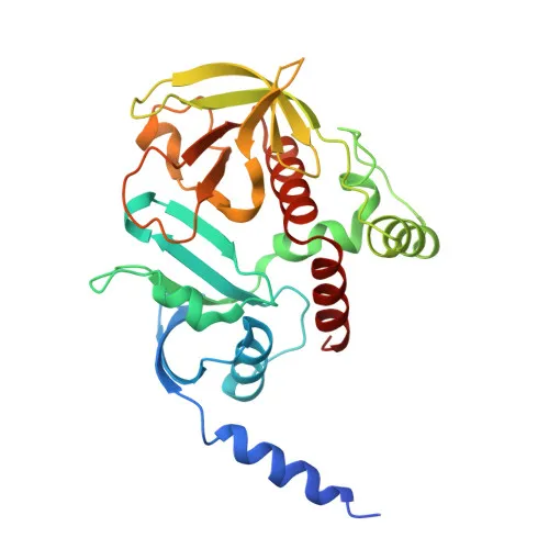
- Post‑translational modifications: Subject to acetylation and phosphorylation; categorized as a phosphoprotein, and its 3D structure has been experimentally determined, underscoring its role in signaling and cytoskeletal remodeling.
Experimental Guidance and Technical Tips
- The polyclonal antibody was raised against a recombinant fusion protein spanning the N‑terminal region (aa 1‑240) of human RAPH1; consider verifying isoform detection specificity using knockdown or isoform‑specific transcript analysis.
- For Western blotting, the predicted molecular weight is ~135 kDa; ensure adequate protein extraction from fresh or well‑preserved samples and consider adding phosphatase inhibitors to preserve phosphorylation states.
- In immunofluorescence/immunocytochemistry (IF/ICC), permeabilization is recommended to access membrane‑associated and cytoskeletal pools; perform appropriate controls to distinguish specific from nonspecific signals.
- Cross‑reactivity with mouse and rat has been reported; when using these species, confirm sequence homology in the immunogen region and optimize conditions for each model system.
CamelBio: Your One-Stop Sourcing Bridge
CamelBio is a dedicated IVD raw‑material partner for diagnostic manufacturers, labs, and research institutes, providing seamless access to validated antibodies, bulk ancillary reagents, and custom sourcing for rare targets. This anti-RAPH1 polyclonal antibody exemplifies our focus on cell signaling and cytoskeletal research tools, and we can supply matched antibody pairs, optimized monoclonal/polyclonal antibodies, and technical consultation to accelerate your assay development from concept to clinic.
REQUEST A QUOTE
Our professional team will reply to you within one business day. Please feel free to contact us!