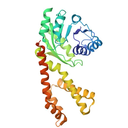
Polyclonal Antibodies
Anti-PYCR2 Polyclonal Antibody for WB, IHC-P, IF/ICC, ELISA - Q96C36
Item Number : CM0018190
Price varies based on specs and customizations
- Application
- WB, IHC-P, IF/ICC, ELISA
- Cross Reactivity
- Human, Mouse, Rat
- Protein Weight
- 34kDa

Shipping:
Contact us to get shipping details Enjoy On-time Dispatch Guarantee.
Why Choose Us
Easy ordering process, quality products, and dedicated support for your business success.
Core Product Specifications and Parameters
| Parameter | Value |
|---|---|
| Product Name | PYCR2 Rabbit pAb |
| Remarks/Alias | HLD10; P5CR2; PYCR2 |
| Species | Human |
| GeneID (Human) | 29920 |
| GeneID | 29920 |
| Immunogen | Recombinant fusion protein containing a sequence corresponding to amino acids 271-320 of human PYCR2 (NP_037460.2). |
| Source | Rabbit |
| Category | Polyclonal Antibodies |
| Application | WB, IHC-P, IF/ICC, ELISA |
| Cross Reactivity | Human, Mouse, Rat |
| SWISS | Q96C36 |
| Protein Weight | 34kDa |
| Shipping | Ice bag |
Biological Background: PYCR2 Function and Localization
- PYCR2 catalyzes the NAD(P)H-dependent reduction of pyrroline-5-carboxylate to L-proline, the final step in proline biosynthesis, with a preference for NADH at physiological concentrations. Related references: PMID:23024808 PMID:2722838 PMID:6894153
- In erythrocytes, PYCR2 primarily generates NADP+, an important cofactor for maintaining reductive potential and protecting against oxidative damage. Related references: PMID:2722838 PMID:6894153
- PYCR2 is involved in the cellular response to oxidative stress. Related references: PMID:25865492
- The enzyme localizes to both the cytoplasm and mitochondria, as evidenced by subcellular fractionation and immunofluorescence studies.
- PYCR2 is detected in erythrocytes at the protein level and is expressed in fetal brain, suggesting roles in both systemic and neural proline metabolism. Related references: PMID:2722838 PMID:6894153
- Post-translational modifications include acetylation and phosphorylation, which may regulate its activity or stability (based on proteomics data).
- Mutations in PYCR2 are linked to leukodystrophy type 10 (HLD10), a severe neurological disorder, underscoring its importance in central nervous system function.
Experimental Guidance and Technical Tips
- The immunogen corresponds to amino acids 271–320 near the C-terminus of human PYCR2. This region is likely exposed in both native and denatured conditions, supporting use in western blotting, immunohistochemistry, and immunofluorescence.
- For western blotting, expect a band around 34 kDa. Validate with positive and negative controls, especially in lysates from human, mouse, or rat tissues.
- For immunohistochemistry (IHC-P), optimal antigen retrieval conditions may need to be determined empirically. Consider using known positive tissues such as fetal brain or erythrocytes.
- In immunofluorescence (IF/ICC), a mitochondrial and cytoplasmic staining pattern is expected; co-staining with organelle markers can confirm localization.
- Since this is a polyclonal antibody, lot-to-lot consistency should be verified, and appropriate storage conditions (shipped on ice bags) should be maintained to preserve activity.
CamelBio: Your One-Stop Sourcing Bridge
CamelBio serves as a comprehensive sourcing partner for IVD raw materials, offering this anti-PYCR2 rabbit polyclonal antibody alongside validated antibody pairs, bulk ancillary reagents, and specialized support for rare-target projects. Our portfolio covers every stage from concept to clinic, ensuring reliable access to critical components for proline metabolism and oxidative stress research. Partner with us to streamline your diagnostic development pipeline.
REQUEST A QUOTE
Our professional team will reply to you within one business day. Please feel free to contact us!