Polyclonal Antibodies
Anti-PTX3 Polyclonal Antibody for WB and ELISA - P26022
Item Number : CM0015684
Price varies based on specs and customizations
- Application
- WB, ELISA
- Cross Reactivity
- Human, Mouse, Rat
- Protein Weight
- 42kDa

Shipping:
Contact us to get shipping details Enjoy On-time Dispatch Guarantee.
Why Choose Us
Easy ordering process, quality products, and dedicated support for your business success.
Core Product Specifications and Parameters
| Parameter | Value |
|---|---|
| Product Name | PTX3 Rabbit pAb |
| Remarks/Alias | TSG-14; TNFAIP5; PTX3 |
| Species | Human |
| Gene ID (Human) | 5806 |
| Gene ID | 5806 |
| Immunogen | Recombinant fusion protein containing a sequence corresponding to amino acids 18-381 of human PTX3 (NP_002843.2) |
| Source | Rabbit |
| Category | Polyclonal Antibodies |
| Application | WB, ELISA |
| Cross Reactivity | Human, Mouse, Rat |
| SWISS | P26022 |
| Protein Weight | 42kDa |
| Shipping | Ice bag |
Biological Background: PTX3 Function and Localization
- Pentraxin-related protein PTX3, also known as tumor necrosis factor-inducible gene 14 protein (TSG-14) or TNF alpha-induced protein 5 (TNFAIP5), belongs to the pentraxin superfamily.
- Functions as a soluble pattern recognition receptor involved in innate immunity, playing a key role in the regulation of resistance to pathogens and inflammatory reactions.
- Participates in the clearance of self-components, such as apoptotic cells, and is implicated in female fertility processes.
- Contains an N-terminal coiled-coil domain and a C-terminal pentraxin domain; classified as a long pentraxin with structural features that support multimerization and ligand binding.
- Secreted glycoprotein with a signal peptide, predicted molecular weight of approximately 42 kDa, and subject to post-translational modifications including N-linked glycosylation and disulfide bond formation.
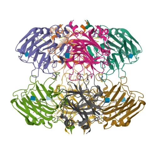
- Based on UniProt keywords, the protein has a known 3D structure and has been identified in proteomic studies, supporting its biochemical characterization.
- Localizes to the extracellular space and is induced in various cell types upon inflammatory stimuli, contributing to tissue homeostasis and repair.
Experimental Guidance and Technical Tips
- The immunogen spans nearly the entire mature human PTX3 protein (amino acids 18–381), suggesting the polyclonal antibody may recognize multiple epitopes and provide robust detection.
- For western blot, validate the antibody in a relevant sample system (e.g., stimulated human, mouse, or rat cell lines or tissues) under reducing and non-reducing conditions; a band near 42 kDa is expected.
- For ELISA, consider using this antibody as a capture or detection reagent; confirm specificity with recombinant PTX3 or relevant biological fluids, and optimize coating and blocking conditions.
- When comparing cross-species reactivity, note that sequence homology and post-translational modifications may influence detection in mouse and rat samples; include appropriate positive and negative controls.
- Store aliquots at –20°C or lower upon receipt and avoid repeated freeze-thaw cycles to maintain antibody integrity.
CamelBio: Your One-Stop Sourcing Bridge
As a specialized IVD raw-material partner, CamelBio connects diagnostic manufacturers and researchers with high-quality antibody reagents like this anti-PTX3 polyclonal antibody. We offer one-stop access to validated antibody pairs, optimized monoclonal and polyclonal antibodies, bulk ancillary reagents, and rare target sourcing—supporting every stage from concept to clinic. Whether you are developing immunoassays for inflammation markers or scaling up production, CamelBio streamlines your raw-material procurement with reliable supply and technical consultation.
REQUEST A QUOTE
Our professional team will reply to you within one business day. Please feel free to contact us!