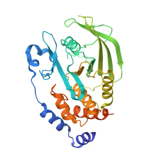
Polyclonal Antibodies
Anti-PTPN2 Polyclonal Antibody (KO Validated) for WB, IF/ICC, ELISA - P17706
Item Number : CM0022048
Price varies based on specs and customizations
- Application
- WB, IF/ICC, ELISA
- Cross Reactivity
- Human, Mouse, Rat
- Protein Weight
- 48kDa

Shipping:
Contact us to get shipping details Enjoy On-time Dispatch Guarantee.
Why Choose Us
Easy ordering process, quality products, and dedicated support for your business success.
Core Product Specifications and Parameters
| Parameter | Value |
|---|---|
| Product Name | [KO Validated] PTPN2 Rabbit pAb |
| Remarks/Alias | PTN2; PTPT; TCPTP; TC-PTP; TCELLPTP; N2 |
| Species | Human |
| GeneID (Human) | 5771 |
| GeneID | 5771 |
| Immunogen | Recombinant fusion protein containing a sequence corresponding to amino acids 1-353 of human PTPN2 (NP_536348.1) |
| Source | Rabbit |
| Category | Polyclonal Antibodies |
| Application | WB, IF/ICC, ELISA |
| Cross Reactivity | Human, Mouse, Rat |
| SWISS | P17706 |
| Protein Weight | 48kDa |
| Shipping | Ice bag |
Biological Background: PTPN2 Function and Localization
- PTPN2, also known as TCPTP (T-cell protein-tyrosine phosphatase), is a non-receptor type tyrosine-specific phosphatase that dephosphorylates receptor protein tyrosine kinases including INSR, EGFR, CSF1R, and PDGFR, as well as non-receptor kinases like JAK1, JAK2, JAK3, and Src family kinases.
- It plays a critical role in immune system development and function, particularly in T-cell receptor signaling through dephosphorylation of FYN and LCK, thereby regulating T-cell differentiation and activation.
- PTPN2 negatively regulates cytokine signaling, including IL-2, interferon, IL-6, and IL-4 pathways, by dephosphorylating STAT1, STAT3, STAT6, and the associated JAK kinases.
- Beyond immunity, PTPN2 is involved in glucose homeostasis by negatively regulating insulin receptor signaling and IL-6-mediated gluconeogenesis, and it also controls cell proliferation by dephosphorylating EGFR, PDGFRB, and MET.
- The protein localizes to multiple subcellular compartments, including the endoplasmic reticulum, ER-Golgi intermediate compartment, nucleus, cytoplasm, and cell membrane.
- Ubiquitously expressed, with isoform 2 being the predominant form; isoform 1 is specifically found in T-cells and placenta.
- Post-translational modifications include phosphorylation and S-nitrosylation, and alternative splicing generates multiple isoforms.
Experimental Guidance and Technical Tips
- The immunogen encompasses amino acids 1–353, covering the catalytic phosphatase domain; this raises the likelihood of recognizing both major isoforms and may be suitable for detecting endogenous PTPN2 in various species.
- For Western blotting, validate the antibody in relevant cell lines or tissues (e.g., Jurkat for T-cell studies) and consider phosphatase inhibitor cocktails during lysis to preserve phosphorylation states.
- In immunofluorescence/ICC, optimize fixation (e.g., 4% PFA) and permeabilization (e.g., 0.1% Triton X-100) to detect PTPN2 in its multiple subcellular locations; include appropriate controls.
- For ELISA, the antibody can be explored as a capture or detection reagent; pairing with a monoclonal antibody may improve specificity, and titration is recommended.
- Cross-reactivity with mouse and rat is indicated; verify performance in your specific model system if using these species.
CamelBio: Your One-Stop Sourcing Bridge
CamelBio is your comprehensive partner for IVD raw materials, supporting diagnostic manufacturers and research labs from concept to clinic. For PTPN2-related immunoassay development—whether in immunology, oncology, or metabolic disease—we offer validated antibody pairs, optimized polyclonal and monoclonal antibodies, bulk ancillary reagents, and custom sourcing for rare targets. Let CamelBio streamline your procurement and accelerate your project’s timeline with reliable, high-quality raw materials.
Product Datasheet
Anti-PTPN2 Polyclonal Antibody (KO Validated) for WB, IF/ICC, ELISA - P17706
REQUEST A QUOTE
Our professional team will reply to you within one business day. Please feel free to contact us!