Monoclonal Antibodies
Anti-PTPIP51/RMDN3 Monoclonal Antibody for WB, ELISA - Q96TC7
Item Number : CM0007346
Price varies based on specs and customizations
- Application
- WB, ELISA
- Cross Reactivity
- Rat
- Protein Weight
- 52kDa

Shipping:
Contact us to get shipping details Enjoy On-time Dispatch Guarantee.
Why Choose Us
Easy ordering process, quality products, and dedicated support for your business success.
Core Product Specifications and Parameters
| Parameter | Value |
|---|---|
| Product Name | PTPIP51/RMDN3 Rabbit mAb |
| Remarks/Alias | RMD3; RMD-3; FAM82C; FAM82A2; ptpip51; PTPIP51/RMDN3 |
| Species | Human |
| GeneID (Human) | 55177 |
| GeneID | 55177 |
| Immunogen | Synthetic peptide corresponding to a sequence within amino acids 200-300 of human PTPIP51/RMDN3 (NP_060615.1) |
| Source | Rabbit |
| Category | Monoclonal Antibodies |
| Application | WB, ELISA |
| Cross Reactivity | Rat |
| SWISS | Q96TC7 |
| Protein Weight | 52kDa |
| Shipping | Ice bag |
Biological Background: PTPIP51/RMDN3 Function and Localization
- PTPIP51/RMDN3, also known as Regulator of microtubule dynamics protein 3, is encoded by the RMDN3 gene (alternative names FAM82A2, FAM82C, and PTPIP51).
- It is a transmembrane protein localized primarily to the mitochondrial outer membrane, but also detected in the cytoplasm, nucleus, and at spindle poles during mitosis.
- Functionally, PTPIP51/RMDN3 is involved in cellular calcium homeostasis regulation, playing a role in maintaining intracellular Ca²⁺ balance.
- The protein may participate in keratinocyte differentiation and apoptosis; its overexpression has been shown to induce apoptosis.
- Tissue expression is prominent in epidermis, seminiferous epithelium, and fast-twitch skeletal muscle fibers, with high levels in suprabasal keratinocytes and differentiating spermatocytes.
- In ciliated epithelia, PTPIP51/RMDN3 associates with microtubular structures of ciliary movement, and it is also found in hippocampal neurons, autonomic ganglion cells, and peripheral nerve axons.
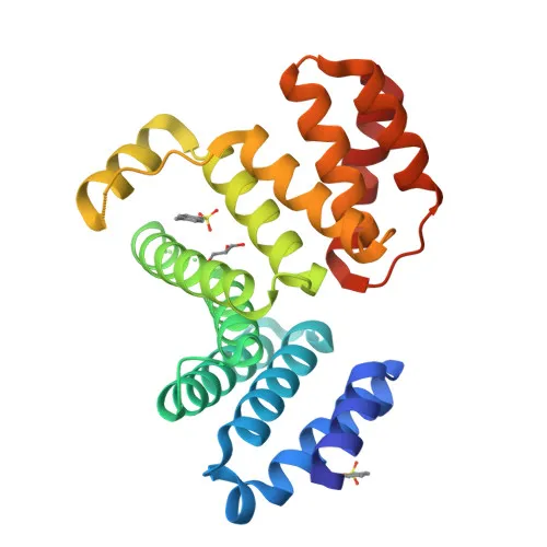
- Post-translational modifications include phosphorylation, and the protein contains a coiled-coil domain and a transmembrane helix, as evidenced by structural studies.
Experimental Guidance and Technical Tips
- The immunogen used is a synthetic peptide within amino acids 200-300, which targets the central region of the protein. For Western blotting, a band at approximately 52 kDa is expected; consider using whole-cell lysates from human or rat cell lines or tissues.
- With cross-reactivity to rat predicted, this antibody may be suitable for comparative studies between human and rat models, but validation in the relevant sample system is recommended.
- For ELISA, the monoclonal nature of the antibody provides high specificity; consider optimizing coating and detection conditions using recombinant protein or peptide as standard.
- Given the protein's localization to mitochondria and cytoskeleton, subcellular fractionation may enhance detection by reducing sample complexity.
- The protein's involvement in apoptosis and differentiation suggests it may be a useful marker in studies of epidermal biology, spermatogenesis, or calcium signaling pathways.
CamelBio: Your One-Stop Sourcing Bridge
CamelBio provides diagnostic manufacturers and research laboratories with comprehensive access to IVD raw materials, technical services, and consulting. For targets like PTPIP51/RMDN3, which is implicated in apoptosis, calcium signaling, and mitochondrial dynamics, CamelBio can deliver validated monoclonal antibodies, bulk ancillary reagents, and support for rare target sourcing. Whether you need antibody pairs for assay development or optimized monoclonal/polyclonal antibodies for research, CamelBio is your trusted partner from concept to clinic.
REQUEST A QUOTE
Our professional team will reply to you within one business day. Please feel free to contact us!