Monoclonal Antibodies
Anti-PTP1B Monoclonal Antibody for WB, IHC-P, ELISA - P18031
Item Number : CM0007166
Price varies based on specs and customizations
- Application
- WB, IHC-P, ELISA
- Cross Reactivity
- Human, Mouse
- Protein Weight
- 50kDa

Shipping:
Contact us to get shipping details Enjoy On-time Dispatch Guarantee.
Why Choose Us
Easy ordering process, quality products, and dedicated support for your business success.
Core Product Specifications and Parameters
| Parameter | Value |
|---|---|
| Product Name | PTP1B Rabbit mAb |
| Remarks/Alias | PTP1B |
| Species | Human |
| GeneID (human) | 5770 |
| GeneID | 5770 |
| Immunogen | Recombinant fusion protein containing a sequence corresponding to amino acids 281-435 of human PTP1B (NP_002818.1). |
| Source | Rabbit |
| Category | Monoclonal Antibodies |
| Application | WB, IHC-P, ELISA |
| Cross Reactivity | Human, Mouse |
| SWISS | P18031 |
| Protein Weight | 50kDa |
| Shipping | Ice bag |
Biological Background: PTP1B Function and Localization
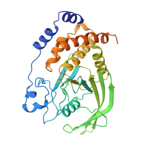
- PTP1B (Tyrosine-protein phosphatase non-receptor type 1) is also known as protein-tyrosine phosphatase 1B (PTP-1B). It is a non-receptor type protein tyrosine phosphatase.
- Acts as a key regulator of the endoplasmic reticulum (ER) unfolded protein response by mediating dephosphorylation and inactivation of the kinase EIF2AK3/PERK.
- Plays a role in signal transduction cascades induced by CKII and p60c-src, and may regulate the EFNA5-EPHA3 signaling pathway involved in cell reorganization and repulsion.
- Modulates the hepatocyte growth factor receptor (MET) signaling pathway through dephosphorylation, thereby influencing downstream cellular responses.
- Located at the endoplasmic reticulum membrane, consistent with its role in ER stress signaling and protein processing.
- Expressed in keratinocytes at the protein level, suggesting a role in skin biology.
- Post-translational modifications include acetylation, oxidation, phosphorylation, and S-nitrosylation, which may regulate its activity and localization.
- Classified under hydrolase, protein phosphatase, and reference proteome; it is a phosphoprotein involved in reversible tyrosine dephosphorylation.
Experimental Guidance and Technical Tips
- The immunogen corresponds to the C-terminal catalytic domain (aa 281-435), which may allow recognition of the active phosphatase region; consider this when designing inhibition or activity assays.
- For Western blotting, the predicted molecular weight is approximately 50 kDa; use appropriate gel percentages and validate detection in your sample of interest.
- In IHC-P, optimize antigen retrieval and antibody dilution for tissue-specific detection, especially in samples with expected expression like keratinocytes.
- For ELISA, pair this antibody with a suitable capture/detection system, and consider cross-reactivity with mouse PTP1B if studying orthologous systems.
- Validate the antibody’s performance in your specific experimental context, as applications listed are based on supplier data.
CamelBio: Your One-Stop Sourcing Bridge
CamelBio streamlines IVD raw-material procurement for diagnostic manufacturers, labs, and research institutes by offering validated antibody pairs, optimized monoclonal/polyclonal antibodies, and bulk ancillary reagents. For studies on tyrosine phosphatase signaling, metabolic disorders, or cancer, CamelBio provides this PTP1B rabbit monoclonal antibody with verified cross-reactivity to human and mouse targets. As your sourcing bridge from concept to clinic, we also assist with rare target raw-material sourcing to accelerate your assay development.
REQUEST A QUOTE
Our professional team will reply to you within one business day. Please feel free to contact us!