Polyclonal Antibodies
Anti-PTK7 Rabbit Polyclonal Antibody for WB, IHC-P, ELISA - Q13308
Item Number : CM0030150
Price varies based on specs and customizations
- Application
- WB, IHC-P, ELISA
- Cross Reactivity
- Human, Mouse, Rat
- Protein Weight
- 118kDa

Shipping:
Contact us to get shipping details Enjoy On-time Dispatch Guarantee.
Why Choose Us
Easy ordering process, quality products, and dedicated support for your business success.
Core Product Specifications and Parameters
| Parameter | Specification |
|---|---|
| Product Name | PTK7 Rabbit pAb |
| Remarks/Alias | CCK4; CCK-4; PTK7 |
| Species | Human |
| GeneID (Human) | 5754 |
| GeneID | 5754 |
| Immunogen | Recombinant fusion protein containing a sequence corresponding to amino acids 811-1070 of human PTK7 (NP_002812.2) |
| Source | Rabbit |
| Category | Polyclonal Antibodies |
| Application | WB, IHC-P, ELISA |
| Cross Reactivity | Human, Mouse, Rat |
| SWISS | Q13308 |
| Protein Weight | 118kDa |
| Shipping | Ice bag |
Biological Background: PTK7 Function and Localization
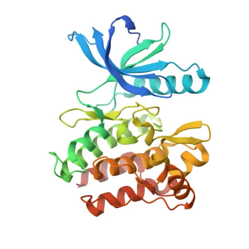
- PTK7 (protein tyrosine kinase 7) is an inactive tyrosine kinase receptor, also known as colon carcinoma kinase 4 (CCK-4), belonging to the receptor tyrosine kinase family. It features an extracellular region with immunoglobulin-like domains, a single-pass transmembrane helix, and a catalytically inactive cytoplasmic kinase domain.
- As a pseudokinase, PTK7 does not bind ATP and lacks phosphotransferase activity, distinguishing it from active kinases.
- PTK7 is a key component of both canonical and non-canonical Wnt signaling pathways, where it regulates multiple cellular processes including cell adhesion, migration, polarity, proliferation, and actin cytoskeleton reorganization. It also plays essential roles in embryogenesis, epithelial tissue organization, and angiogenesis. Related references: PMID:24107129 PMID:32619402
- Localized primarily at the cell membrane and cell junctions, PTK7 mediates cell-cell and cell-matrix interactions.
- PTK7 exhibits high expression in lung, liver, pancreas, kidney, placenta, and melanocytes; weak expression in thyroid gland, ovary, brain, heart, and skeletal muscle; and is absent from colon tissue.
- Post-translational modifications include glycosylation and phosphorylation, and the protein contains disulfide bonds and a signal peptide.
Experimental Guidance and Technical Tips
- For Western blotting, the expected molecular weight is approximately 118 kDa. Use appropriate gel percentage and transfer conditions for large proteins.
- For IHC-P, PTK7 is highly expressed in lung, placenta, and kidney tissues; include these as positive controls and optimize antigen retrieval protocols.
- The antibody cross-reacts with mouse and rat PTK7; consider validating signal specificity in the relevant species and tissue context.
- As a polyclonal antibody, lot-to-lot variability may occur; perform pilot experiments and titrate the antibody for each application.
CamelBio: Your One-Stop Sourcing Bridge
CamelBio provides diagnostic manufacturers and research institutions with one-stop access to IVD raw materials, including validated antibody pairs, optimized monoclonal/polyclonal antibodies, and bulk ancillary reagents. This PTK7 rabbit polyclonal antibody supports Wnt signaling and cell adhesion research, and CamelBio facilitates seamless sourcing from concept to clinic. For rare target raw-material requirements, our team offers tailored procurement solutions to accelerate your development pipelines.
REQUEST A QUOTE
Our professional team will reply to you within one business day. Please feel free to contact us!