Polyclonal Antibodies
Anti-PSTPIP1 Rabbit Polyclonal Antibody for WB, IHC-P, ELISA - O43586
Item Number : CM0026494
Price varies based on specs and customizations
- Application
- WB, IHC-P, ELISA
- Cross Reactivity
- Human, Mouse, Rat
- Protein Weight
- 48kDa

Shipping:
Contact us to get shipping details Enjoy On-time Dispatch Guarantee.
Why Choose Us
Easy ordering process, quality products, and dedicated support for your business success.
Core Product Specifications and Parameters
| Parameter | Value |
|---|---|
| Product Name | PSTPIP1 Rabbit pAb |
| Remarks/Alias | H-PIP; PAPAS; CD2BP1; PSTPIP; CD2BP1L; CD2BP1S; PSTPIP1 |
| Species | Human |
| GeneID (Human) | 9051 |
| Immunogen | Recombinant protein |
| Source | Rabbit |
| Category | Polyclonal Antibodies |
| Application | WB, IHC-P, ELISA |
| Cross Reactivity | Human, Mouse, Rat |
| SWISS | O43586 |
| Protein Weight | 48kDa |
| Shipping | Ice bag |
Biological Background: PSTPIP1 Function and Localization
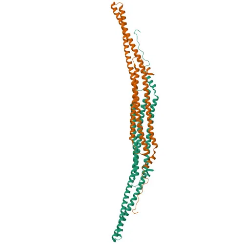
- PSTPIP1 (Proline-serine-threonine phosphatase-interacting protein 1), also known as CD2BP1 and H-PIP, is a scaffold protein containing a coiled-coil domain and an SH3 domain. Alternative splicing produces multiple isoforms (CD2BP1L, CD2BP1S).
- It plays a key role in actin cytoskeleton regulation by bridging interactions between ABL1 and PTPN18, leading to ABL1 dephosphorylation, and by acting as a scaffold between PTPN12 and WAS, facilitating WAS dephosphorylation.
- In T-cells, PSTPIP1 functions downstream of CD2 and CD2AP to recruit WAS to the T-cell:APC contact site, promoting actin polymerization required for synapse induction during T-cell activation.
- PSTPIP1 is involved in innate immunity and inflammatory responses: it is recruited to inflammasomes by MEFV, induces pyroptosome formation through PYCARD oligomerization, and facilitates MEFV-PYCARD binding, leading to inflammatory apoptosis.
- It down-regulates CD2-stimulated adhesion by coupling PTPN12 to CD2, and regulates endocytosis and cell migration in neutrophils.
- Subcellular localization includes cytoplasm, cell membrane, uropodium, lamellipodium, cleavage furrow, perinuclear region, and cytoskeleton, consistent with its roles in cell adhesion and migration.
- Tissue specificity: highly expressed in peripheral blood leukocytes, granulocytes, monocytes (particularly T-cells and NK cells), and spleen; weakly expressed in thymus, small intestine, lung, and placenta.
- Post-translational modifications: classified as a phosphoprotein (UniProt keyword), suggesting regulation by phosphorylation.
Experimental Guidance and Technical Tips
- The immunogen corresponds to amino acids 157-416 of human PSTPIP1, a region encompassing functionally important domains. For Western blot, a band around 48 kDa is expected, but validate in your sample system as post-translational modifications or isoforms may produce additional bands.
- For IHC-P, consider antigen retrieval optimization and test on positive control tissues such as spleen or leukocyte-rich samples. Cross-reactivity with mouse and rat allows comparative studies; verify species-specific reactivity in your model.
- For ELISA, determine the optimal coating and detection conditions. Polyclonal nature may reduce sensitivity to epitope loss, but titrate to minimize background.
CamelBio: Your One-Stop Sourcing Bridge
CamelBio supports diagnostic manufacturers, labs, and research institutes as a comprehensive IVD raw-material sourcing partner. We supply validated antibody pairs, optimized monoclonal and polyclonal antibodies, bulk ancillary reagents, and rare target raw materials for immunoassay development. For immunology and inflammation research, CamelBio bridges your project needs with high-quality reagents like anti-PSTPIP1 antibodies to accelerate your development pipeline.
Product Datasheet
Anti-PSTPIP1 Rabbit Polyclonal Antibody for WB, IHC-P, ELISA - O43586
REQUEST A QUOTE
Our professional team will reply to you within one business day. Please feel free to contact us!