Polyclonal Antibodies
Anti-PRKCSH Polyclonal Antibody for WB and ELISA - P14314
Item Number : CM0029663
Price varies based on specs and customizations
- Application
- WB, ELISA
- Cross Reactivity
- Human, Mouse, Rat
- Protein Weight
- 59kDa

Shipping:
Contact us to get shipping details Enjoy On-time Dispatch Guarantee.
Why Choose Us
Easy ordering process, quality products, and dedicated support for your business success.
Core Product Specifications and Parameters
| Parameter | Value |
|---|---|
| Product Name | PRKCSH Rabbit pAb |
| Remarks/Alias | GIIB; PCLD; PLD1; G19P1; PCLD1; PKCSH; AGE-R2; VASAP-60; PRKCSH |
| Species | Human |
| GeneID | 5589 |
| Immunogen | Recombinant fusion protein containing a sequence corresponding to amino acids 130-275 of human PRKCSH (P14314) |
| Source | Rabbit |
| Category | Polyclonal Antibodies |
| Application | WB, ELISA |
| Cross Reactivity | Human, Mouse, Rat |
| SWISS | P14314 |
| Protein Weight | 59kDa |
| Shipping | Ice bag |
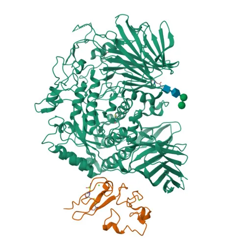
Biological Background: PRKCSH Function and Localization
- PRKCSH encodes glucosidase 2 subunit beta (also known as 80K-H protein or PKCSH), a key regulator of N-glycan processing.
- It serves as the regulatory subunit of glucosidase II, which cleaves the two innermost α-1,3-linked glucose residues from Glc₂Man₉GlcNAc₂ oligosaccharide precursors of immature glycoproteins. Related references: PMID:10929008
- This glucose trimming is critical for glycoprotein folding and ER quality control, ensuring only properly folded proteins exit the ER.
- PRKCSH is required for efficient PKD1/polycystin-1 biogenesis and trafficking to the primary cilium plasma membrane.
- The protein localizes on the endoplasmic reticulum membrane.
- It contains calcium-binding domains and is itself a glycoprotein; published phosphoproteomic data identify multiple phosphorylation sites.
- Mutations in PRKCSH cause autosomal dominant polycystic liver disease (PCLD).
- Alternative splicing produces multiple transcript variants, adding functional diversity.
Experimental Guidance and Technical Tips
- The immunogen corresponds to a central region (aa 130–275) of human PRKCSH; the polyclonal antibody is expected to recognize both full-length and potential splice variants or fragments containing this epitope.
- For Western blot, a major band near 59 kDa is anticipated. Reducing and denaturing conditions are recommended; pilot experiments should confirm specificity.
- In ELISA, recombinant PRKCSH protein or cell lysates from human, mouse, or rat origin can be used as capture/detection reagents. Optimize coating concentrations and detection conditions.
- Reported cross-reactivity with mouse and rat suggests the epitope is conserved; however, sequence alignment and preliminary testing are advised when working with non-human samples.
- As with any polyclonal antibody, lot-to-lot variability may occur. Titrate the antibody in the specific assay system and include appropriate positive/negative controls.
CamelBio: Your One-Stop Sourcing Bridge
CamelBio positions this anti-PRKCSH polyclonal antibody within a comprehensive portfolio of IVD raw materials, serving diagnostic manufacturers and research laboratories focused on glycoprotein processing, polycystic kidney disease, and ER-associated pathways. To accelerate assay development, CamelBio can supply validated antibody pairs, optimized monoclonal and polyclonal antibodies, bulk ancillary reagents, and support for rare-target raw-material sourcing. Reach out to explore how CamelBio’s integrated procurement services can streamline your diagnostic pipeline from concept to clinical application.
REQUEST A QUOTE
Our professional team will reply to you within one business day. Please feel free to contact us!