Monoclonal Antibodies
Anti-PRK2/PKN2 Monoclonal Antibody for WB and ELISA - Q16513
Item Number : CM0002768
Price varies based on specs and customizations
- Application
- WB, ELISA
- Cross Reactivity
- Human, Mouse, Rat
- Protein Weight
- 112kDa

Shipping:
Contact us to get shipping details Enjoy On-time Dispatch Guarantee.
Why Choose Us
Easy ordering process, quality products, and dedicated support for your business success.
Core Product Specifications and Parameters
| Specification | Details |
|---|---|
| Product Name | PRK2/PKN2 Rabbit mAb |
| Remarks/Alias | PAK2; PRK2; STK7; Pak-2; PRKCL2; PRO2042; PRK2/PKN2 |
| Species | Human |
| GeneID (Human) | 5586 |
| GeneID | 5586 |
| Immunogen | Synthetic Peptide |
| Source | Rabbit |
| Category | Monoclonal Antibodies |
| Application | WB, ELISA |
| Cross Reactivity | Human, Mouse, Rat |
| SWISS | Q16513 |
| Protein Weight | 112kDa |
| Shipping | Ice bag |
Biological Background: PRK2/PKN2 Function and Localization
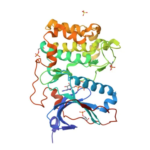
- PRK2/PKN2 (also known as PKN gamma, PAK2, PRKCL2) is a serine/threonine-protein kinase belonging to the protein kinase C (PKC) family. It functions as a Rho/Rac effector involved in various signal transduction pathways.
- The kinase participates in regulating cell cycle progression, specifically the G2/M transition and cytokinesis, as well as actin cytoskeleton assembly, cell migration, adhesion, and tumor cell invasion.
- It directly targets RhoA and is required for the maturation of primordial junctions into apical junction formation in bronchial epithelial cells, and it stimulates FYN kinase activity for skin cell-cell adhesion during keratinocyte differentiation.
- Subcellularly, PRK2/PKN2 localizes to the cytoplasm, nucleus, membrane, lamellipodia, cleavage furrow, midbody, and cell junctions, reflecting its dynamic roles.
- A key function is mediating Rho protein-induced transcriptional activation via the c-fos serum response factor (SRF), and it also phosphorylates HDAC5 to impair its nuclear import, influencing transcription regulation.
- PRK2/PKN2 negatively regulates ciliogenesis. Related references: PMID:27104747
- Additionally, during microbial infection, it phosphorylates HCV NS5B, stimulating HCV RNA replication.
- The protein is ubiquitously expressed and found in numerous tumor cell lines, especially bladder tumor cells, and it inhibits Akt pro-survival kinase activity, linking it to apoptosis pathways.
Experimental Guidance and Technical Tips
- The immunogen is a synthetic peptide from the N-terminal region (amino acids 1–100). The antibody likely recognizes the N-terminal domain; consider its utility in probing N-terminal interactions or modifications.
- For Western blotting, a band at approximately 112 kDa is expected. Validate the antibody using positive control lysates from human cell lines (e.g., HeLa, bladder tumor cells) with confirmed endogenous expression.
- In ELISA, ensure optimal coating with the immunogen peptide or purified recombinant protein. Titrate the antibody to achieve desired sensitivity and specificity.
- Cross-reactivity with mouse and rat is reported; confirm sequence homology in the target regions of these species if using for non-human samples.
CamelBio: Your One-Stop Sourcing Bridge
CamelBio is a dedicated one-stop sourcing partner for IVD raw materials, supporting diagnostic manufacturers and research labs from concept to clinic. For projects focused on PRK2/PKN2 signaling in cancer, cytoskeletal dynamics, or host–pathogen interactions, we offer validated antibody pairs, recombinant proteins, and bulk ancillary reagents. Contact us to access rare-target raw materials and accelerate your assay development with reliable supply solutions.
REQUEST A QUOTE
Our professional team will reply to you within one business day. Please feel free to contact us!