Polyclonal Antibodies
Anti-PMAIP1 (NOXA/APR) Polyclonal Antibody for WB, IF/ICC, ELISA - Q13794
Item Number : CM0025355
Price varies based on specs and customizations
- Application
- WB, IF/ICC, IF-P, ELISA
- Cross Reactivity
- Human, Mouse, Rat
- Protein Weight
- 6kDa

Shipping:
Contact us to get shipping details Enjoy On-time Dispatch Guarantee.
Why Choose Us
Easy ordering process, quality products, and dedicated support for your business success.
Core Product Specifications and Parameters
| Parameter | Value |
|---|---|
| Product Name | PMAIP1 Rabbit pAb |
| Remarks/Alias | APR, NOXA, PMAIP1 |
| Species | Human |
| GeneID (Human) | 5366 |
| GeneID | 5366 |
| Immunogen | Synthetic peptide: A synthetic peptide corresponding to a sequence within amino acids 1-54 of human PMAIP1 (NP_066950.1). |
| Source | Rabbit |
| Category | Polyclonal Antibodies |
| Application | WB, IF/ICC, IF-P, ELISA |
| Cross Reactivity | Human, Mouse, Rat |
| SWISS | Q13794 |
| Protein Weight | 6kDa |
| Shipping | Ice bag |
Biological Background: PMAIP1 Function and Localization
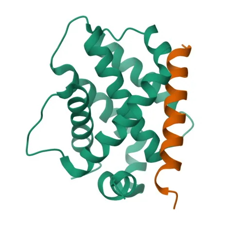
- PMAIP1, also called NOXA or APR, is a pro-apoptotic protein that promotes caspase activation and mitochondrial apoptosis.
- It induces mitochondrial membrane permeabilization and the release of apoptogenic factors from mitochondria.
- PMAIP1 mediates p53/TP53-dependent apoptosis, particularly in response to DNA damage such as radiation exposure.
- The protein functions primarily by binding and neutralizing the anti-apoptotic protein MCL1, thereby freeing pro-apoptotic BAK1 and BIM.
- It directly competes with BAK1 and BIM/BCL2L11 for binding to MCL1, displacing them to trigger apoptosis.
- PMAIP1 is localized to the mitochondrion, consistent with its role in intrinsic apoptosis.
- It is highly expressed in adult T-cell leukemia cell lines, linking it to hematological malignancy regulation.
- Keywords: Alternative splicing, 3D-structure, Apoptosis, Mitochondrion, Reference proteome.
Experimental Guidance and Technical Tips
- The immunogen corresponds to the N-terminal region (aa 1-54); consider this when planning epitope-specific experiments.
- For Western blot, PMAIP1 is a small protein (~6 kDa); ensure adequate membrane pore size and transfer conditions to retain low-molecular-weight proteins.
- In immunocytochemistry/immunofluorescence, mitochondrial localization can be validated by co-staining with mitochondrial markers.
- For ELISA, the antibody can be used as capture or detection reagent; titration is recommended to optimize signal-to-noise ratio.
- Because PMAIP1 is stress-inducible, consider appropriate cell treatments (e.g., DNA damage, serum starvation) to upregulate expression in experimental models.
CamelBio: Your One-Stop Sourcing Bridge
CamelBio provides diagnostic manufacturers and research labs with streamlined access to IVD raw materials, from monoclonal and polyclonal antibodies to ancillary reagents. This PMAIP1 antibody supports apoptosis research and mitochondrial function studies, and we can supply complementary tools such as validated antibody pairs, optimized secondary antibodies, and bulk reagents for large-scale assay development. Our one-stop sourcing model accelerates your workflow from concept to clinic, including rare target material sourcing.
Product Datasheet
Anti-PMAIP1 (NOXA/APR) Polyclonal Antibody for WB, IF/ICC, ELISA - Q13794
REQUEST A QUOTE
Our professional team will reply to you within one business day. Please feel free to contact us!