Polyclonal Antibodies
Anti-PLK3 Polyclonal Antibody for WB, ELISA - Q9H4B4
Item Number : CM0025665
Price varies based on specs and customizations
- Application
- WB, ELISA
- Cross Reactivity
- Human, Mouse, Rat
- Protein Weight
- 72kDa

Shipping:
Contact us to get shipping details Enjoy On-time Dispatch Guarantee.
Why Choose Us
Easy ordering process, quality products, and dedicated support for your business success.
Core Product Specifications and Parameters
| Parameter | Value |
|---|---|
| Product Name | PLK3 Rabbit pAb |
| Remarks/Alias | CNK; FNK; PRK; PLK-3; PLK3 |
| Species | Human |
| GeneID (Human) | 1263 |
| GeneID | 1263 |
| Immunogen | Recombinant fusion protein containing a sequence corresponding to amino acids 487-646 of human PLK3 (NP_004064.2) |
| Source | Rabbit |
| Category | Polyclonal Antibodies |
| Application | WB, ELISA |
| Cross Reactivity | Human, Mouse, Rat |
| SWISS | Q9H4B4 |
| Protein Weight | 72kDa |
| Shipping | Ice bag |
Biological Background: PLK3 Function and Localization
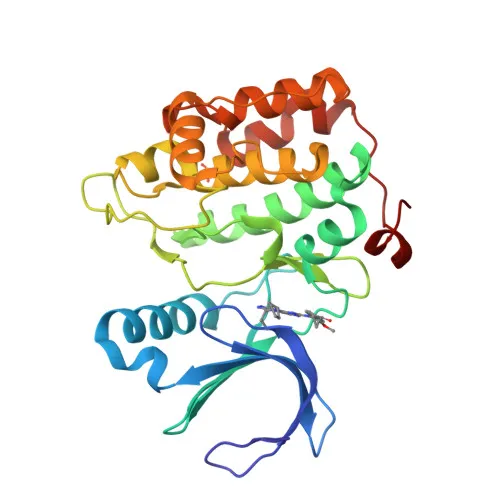
- Protein identity and family: Serine/threonine-protein kinase PLK3 (also known as cytokine-inducible serine/threonine-protein kinase, FGF-inducible kinase, proliferation-related kinase) is a member of the polo-like kinase family. It contains a catalytic domain and POLO box domains that mediate substrate recognition through phosphorylation-dependent binding.
- Cell cycle regulation: PLK3 is required for entry into S phase and cytokinesis. It regulates the G2 checkpoint and mitotic progression by phosphorylating CDC25C and BCL2L1, influencing cell cycle transitions.
- Stress response and DNA damage: PLK3 is rapidly activated upon stress stimuli such as ionizing radiation, reactive oxygen species (ROS), hyperosmotic stress, UV irradiation, and hypoxia. It phosphorylates p53/TP53 to promote apoptosis, CHEK2 to enforce the G2/M checkpoint, and p73/TP73 to inhibit its pro-apoptotic functions.
- Key phosphorylation targets: PLK3 substrates include ATF2, BCL2L1, CDC25A, CDC25C, CHEK2, HIF1A, JUN, p53/TP53, p73/TP73, PTEN, TOP2A, and VRK1. These interactions mediate its roles in checkpoint control, apoptosis, and Golgi dynamics.
- Golgi disassembly: PLK3 participates in Golgi fragmentation during mitosis via a MEK1/MAP2K1-dependent pathway by phosphorylating VRK1 at the Golgi apparatus, ensuring proper organelle partitioning.
- Subcellular localization: PLK3 exhibits dynamic localization to the cytoplasm, nucleus, nucleolus, Golgi apparatus, and centrosome, consistent with its multi-faceted functions in cell cycle and stress signaling.
- Tissue and differentiation expression: Transcripts are highly detected in placenta, lung, skeletal muscle, heart, pancreas, ovaries, and kidney; weakly in liver and brain. In hematopoietic cells, PLK3 is strongly and specifically expressed in terminally differentiated macrophages. Expression is down-regulated in primary lung tumors.
- Post‑translational modifications and keywords: PLK3 is classified as a phosphoprotein (proteomics identification) with ATP‑binding, serine/threonine‑protein kinase activity. UniProt keywords include apoptosis, cell cycle, DNA damage, Golgi apparatus, and reference proteome.
Experimental Guidance and Technical Tips
- The immunogen spans amino acids 487–646 (C‑terminal region), covering part of the second POLO box domain. This region may offer specificity within the PLK family; cross‑reactivity with PLK1 or PLK2 should be confirmed experimentally if required.
- For Western blot (WB), the predicted molecular weight is ~72 kDa. Validate detection in the intended species (human, mouse, rat) and sample type. Use appropriate loading controls and consider siRNA‑mediated knockdown or knockout lysates to verify band identity.
- ELISA applications can be optimized by testing coating conditions and antibody dilutions; the antibody is supplied as a rabbit polyclonal, suitable for standard indirect ELISA protocols.
- To study stress‑induced pathways, treat cells with DNA‑damaging agents (e.g., etoposide, UV), ROS inducers (e.g., H₂O₂), or hypoxia mimetics (e.g., CoCl₂) and confirm PLK3 activation or expression changes by WB.
- For immunofluorescence or immunohistochemistry, users should establish fixation and permeabilization conditions compatible with the detected subcellular compartments (cytoplasmic, nuclear, Golgi). Note that endogenous PLK3 levels may be low in some cell types; enrichment or signal amplification may be necessary.
CamelBio: Your One-Stop Sourcing Bridge
CamelBio offers this PLK3 rabbit polyclonal antibody as part of an extensive portfolio of IVD raw materials, supporting manufacturers and research institutes in accelerating assay development for cell cycle and oncology biomarkers. With robust capabilities in validated antibody pairs, optimized monoclonal/polyclonal antibodies, bulk ancillary reagents, and rare‑target sourcing, CamelBio streamlines procurement from early‑stage concept to clinical application. Contact us to explore custom antibody services and integrated supply solutions tailored to your diagnostic project needs.
REQUEST A QUOTE
Our professional team will reply to you within one business day. Please feel free to contact us!