Monoclonal Antibodies
Anti-Plexin A1 Rabbit Monoclonal Antibody for WB and ELISA - Q9UIW2
Item Number : CM0011142
Price varies based on specs and customizations
- Application
- WB, ELISA
- Cross Reactivity
- Human, Mouse, Rat
- Protein Weight
- 211kDa

Shipping:
Contact us to get shipping details Enjoy On-time Dispatch Guarantee.
Why Choose Us
Easy ordering process, quality products, and dedicated support for your business success.
Core Product Specifications and Parameters
| Parameter | Value |
|---|---|
| Product Name | Plexin A1 Rabbit mAb |
| Remarks/Alias | NOV; NOVP; PLXN1; DWOPNED; PLEXIN-A1; Plexin A1 |
| Species | Human |
| GeneID (Human) | 5361 |
| GeneID | 5361 |
| Immunogen | Synthetic peptide corresponding to a sequence within amino acids 100-200 of human Plexin A1 (Q9UIW2) |
| Source | Rabbit |
| Category | Monoclonal Antibodies |
| Application | WB, ELISA |
| Cross Reactivity | Human, Mouse, Rat |
| SWISS | Q9UIW2 |
| Protein Weight | 211kDa |
| Shipping | Ice bag |
Biological Background: Plexin A1 Function and Localization
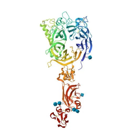
- Plexin-A1 (PLXNA1, semaphorin receptor NOV) is a single-pass type I membrane protein and member of the plexin receptor family, characterized by a large sema domain, multiple PSI domains, IPT repeats, and a conserved cytoplasmic tail.
- It acts as a coreceptor for class 3 semaphorins (SEMA3A, SEMA3C, SEMA3F) and SEMA6D, forming a holoreceptor complex with neuropilins to mediate semaphorin-induced signaling.
- Upon ligand binding, the cytoplasmic domain of plexin-A1 triggers intracellular cascades that regulate cytoskeletal dynamics, contributing to its roles in axon guidance, cell migration, and invasive growth.
- It functions as a coreceptor of TREM2 for SEMA6D in dendritic cells, playing a role in immune response generation and skeletal homeostasis.
- Subcellular localization is primarily at the cell membrane.
- Tissue expression was detected in fetal brain, lung, liver, and kidney.
- Key structural features include a signal peptide, a single-pass transmembrane helix, disulfide bonds, glycosylation sites, and coiled-coil regions; it is annotated with repeats, a transmembrane domain, and an intracellular tail.
- Disease-associated variants in PLXNA1 have been linked to intellectual disability.
Experimental Guidance and Technical Tips
- The immunogen spans amino acids 100–200, which likely lies in the extracellular domain of Plexin-A1; therefore, the antibody may recognize the native protein on intact cells, but denaturing conditions (e.g., SDS-PAGE preparation) could influence epitope accessibility. Validate detection in your relevant sample system.
- For Western blot, the predicted molecular weight is approximately 211 kDa. Use a high-molecular-weight prestained marker and prolonged electrophoresis for optimal separation. Reducing agents (DTT or β-mercaptoethanol) are recommended to disrupt potential disulfide bonds.
- For ELISA, both direct and capture formats may be feasible. Empirical titration of the antibody is essential to determine the optimal working concentration, as no quantitative dilution data are provided.
CamelBio: Your One-Stop Sourcing Bridge
CamelBio offers diagnostic manufacturers and research laboratories a single-source solution for IVD raw materials, including expertly developed monoclonal antibodies such as this anti-Plexin A1 rabbit mAb. We provide validated antibody pairs, bulk ancillary reagents, and tailored sourcing for rare targets, supporting your axon guidance or semaphorin signaling research from concept to clinic. Contact us to discuss your project's specific requirements and procurement needs.
REQUEST A QUOTE
Our professional team will reply to you within one business day. Please feel free to contact us!