Polyclonal Antibodies
Anti-PKD2 Rabbit Polyclonal Antibody for WB, ELISA - Q13563
Item Number : CM0025753
Price varies based on specs and customizations
- Application
- WB, ELISA
- Cross Reactivity
- Human, Mouse, Rat
- Protein Weight
- 110kDa

Shipping:
Contact us to get shipping details Enjoy On-time Dispatch Guarantee.
Why Choose Us
Easy ordering process, quality products, and dedicated support for your business success.
Core Product Specifications and Parameters
| Parameter | Value |
|---|---|
| Product Name | PKD2 Rabbit pAb |
| Remarks/Alias | PC2; PKD4; Pc-2; APKD2; TRPP2; PKD2 |
| Species | Human |
| GeneID (Human) | 5311 |
| GeneID | 5311 |
| Immunogen | Recombinant fusion protein containing a sequence corresponding to amino acids 879-968 of human PKD2 (NP_000288.1) |
| Source | Rabbit |
| Category | Polyclonal Antibodies |
| Application | WB, ELISA |
| Cross Reactivity | Human, Mouse, Rat |
| SWISS | Q13563 |
| Protein Weight | 110kDa |
| Shipping | Ice bag |
Biological Background: Polycystin-2 Function and Localization
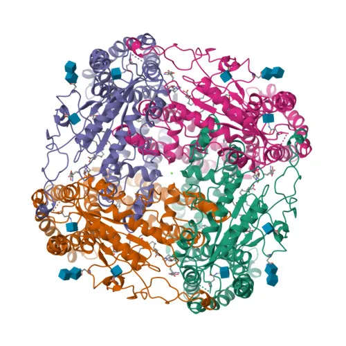
- PKD2, also known as TRPP2 or polycystin-2, is a transmembrane protein encoded by the PKD2 gene, associated with autosomal dominant polycystic kidney disease.
- Functions as a nonselective cation channel, forming homotetramers or heteromers with PKD1; the PKD1/PKD2 heteromer exhibits higher Ca2+ permeability than homomeric PKD2 channels. Related references: PMID:11854751 PMID:11991947 PMID:31441214
- In the primary cilium, PKD2 acts as a cation channel with a preference for monovalent cations and low selectivity for Ca2+, involved in fluid-flow mechanosensation. Related references: PMID:27071085
- Together with TRPV4, forms mechano- and thermosensitive channels in cilium. Related references: PMID:18695040
- Subcellular localization includes the cilium membrane, endoplasmic reticulum membrane, cell membrane, and cytoplasmic vesicles, reflecting its diverse regulatory roles.
- Tissue expression is prominent in fetal and adult kidney, ovary, testis, and small intestine, with protein detected in renal tubules and placental syncytiotrophoblasts. Related references: PMID:10770959 PMID:26269590
- Post-translational modifications include glycosylation and phosphorylation, as indicated by keywords such as glycoprotein and phosphoprotein.
Experimental Guidance and Technical Tips
- The immunogen corresponds to the C-terminal cytoplasmic domain (aa 879–968) of human PKD2, predicted to be exposed; this region is suitable for generating antibodies reactive with the full-length protein under denaturing conditions.
- For western blotting, PKD2 has an expected molecular weight of ~110 kDa (also reported as 109.7 kDa); use a 6–8% SDS-PAGE gel and consider PVDF membranes for efficient transfer of high-molecular-weight proteins.
- The antibody is indicated to cross-react with mouse and rat; validate specificity in your sample system, ideally using kidney tissue lysates or PKD2-overexpressing cells as positive controls.
- When performing ELISA, titrate the antibody to determine optimal coating and detection concentrations; the polyclonal nature may provide robust signal for antigen capture or detection workflows.
CamelBio: Your One-Stop Sourcing Bridge
CamelBio is a dedicated IVD raw-material supply partner, offering this anti-PKD2 polyclonal antibody alongside a comprehensive portfolio of validated antibody pairs, optimized monoclonal/polyclonal antibodies, bulk ancillary reagents, and rare target sourcing. Our expertise in molecular biology and diagnostics accelerates your development of assays for polycystin-2-related research and polycystic kidney disease applications. From concept to clinic, we streamline procurement so you can focus on innovation.
REQUEST A QUOTE
Our professional team will reply to you within one business day. Please feel free to contact us!