Polyclonal Antibodies
Anti-PKACα Rabbit Polyclonal Antibody for WB and ELISA - P17612
Item Number : CM0022591
Price varies based on specs and customizations
- Application
- WB, ELISA
- Cross Reactivity
- Human, Mouse, Rat
- Protein Weight
- 39kDa/40kDa

Shipping:
Contact us to get shipping details Enjoy On-time Dispatch Guarantee.
Why Choose Us
Easy ordering process, quality products, and dedicated support for your business success.
Core Product Specifications and Parameters
| Parameter | Value |
|---|---|
| Product Name | PKACα Rabbit pAb |
| Remarks/Alias | PRKACA, PKACA, PPNAD4, cAMP-dependent protein kinase catalytic subunit alpha, PKACα |
| Species | Human |
| GeneID | 5566 (Human) |
| Immunogen | A synthetic peptide corresponding to a sequence within amino acids 251-351 of human PKACα (NP_002721.1). |
| Source | Rabbit |
| Category | Polyclonal Antibodies |
| Application | WB, ELISA |
| Cross Reactivity | Human, Mouse, Rat |
| SWISS | P17612 |
| Protein Weight | 39kDa/40kDa |
| Shipping | Ice bag |
Biological Background: PKACα Function and Localization
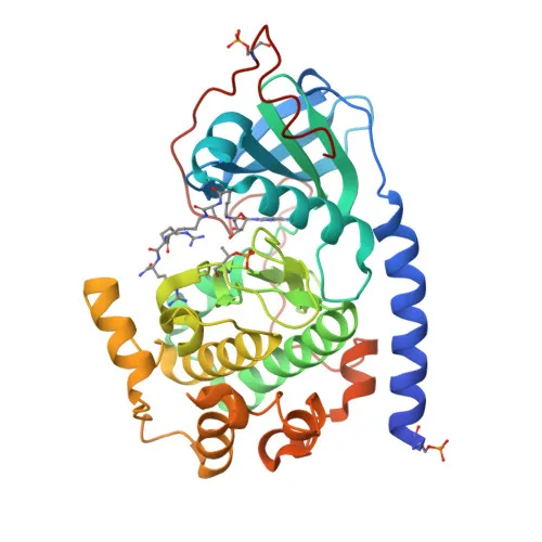
- PKACα (PRKACA) is the catalytic subunit alpha of cAMP-dependent protein kinase. Two isoforms exist: ubiquitous isoform 1 and sperm-specific isoform 2 enriched in pachytene spermatocytes.
- It phosphorylates numerous cytoplasmic and nuclear substrates, including CDC25B, ABL1, NFKB1, histone H1.4, and VASP, regulating cell cycle, transcription, and motility. Related references: PMID:15642694, PMID:15905176, PMID:16387847
- In platelets, PKACα complexes with NF-κB and IκBα to enforce quiescence; agonist stimulation dissociates the complex, freeing PKACα to phosphorylate VASP and promote aggregation. Related references: PMID:15642694, PMID:20356841
- PKACα inhibits smoothened signaling by phosphorylating GLI transcription factors (GLI1/2/3), triggering their processing and blocking target gene expression in the absence of hedgehog ligands. Related references: PMID:16705181
- Sequestration of PKACα by the Smoothened receptor at the cell membrane prevents GLI phosphorylation, allowing pathway activation. Related references: PMID:36202993, PMID:39138140
- PKACα potentiates RYR2 calcium channel activity, altering calcium release dynamics and contributing to store overload-induced Ca²⁺ release. Related references: PMID:17693412
- It negatively regulates mTORC1 by phosphorylating RPTOR, linking cAMP signaling to nutrient sensing. Related references: PMID:31112131
- Subcellularly, PKACα is found in the cytoplasm, nucleus, cell membrane, mitochondria, and cell projections (cilia/flagella), reflecting its multifaceted functions.
Experimental Guidance and Technical Tips
- The immunogen corresponds to a conserved region (amino acids 251-351) within the catalytic domain; cross-reactivity with other PKA catalytic subunits (PRKACB, PRKACG) is possible, so verify specificity in your system.
- In Western blot, a major band at ~39–40 kDa is anticipated; additional bands may represent isoforms or degradation products. Use appropriate positive and negative controls.
- For ELISA, titrate both coating antigen and antibody to optimize signal-to-noise ratio; consider using recombinant PKACα fragment as a standard.
- Given its role in multiple signaling pathways, treat samples with phosphatase inhibitors to preserve phosphorylation-dependent interactions if studying such contexts.
CamelBio: Your One-Stop Sourcing Bridge
CamelBio provides diagnostic manufacturers, labs, and research institutes with one-stop access to IVD raw materials, technical services, and consulting—covering every stage from concept to clinic. This anti-PKACα antibody supports research into cAMP/PKA signaling, kinase activity, and related disorders such as endocrine and cardiovascular diseases. Our portfolio includes validated antibody pairs, optimized monoclonal/polyclonal antibodies, bulk ancillary reagents, and rare target raw-material sourcing to accelerate your development.
REQUEST A QUOTE
Our professional team will reply to you within one business day. Please feel free to contact us!