Polyclonal Antibodies
Anti-PIK3CA Polyclonal Antibody for WB, IHC-P, IF/ICC, ELISA - P42336
Item Number : CM0013294
Price varies based on specs and customizations
- Application
- WB, IHC-P, IF/ICC, ELISA
- Cross Reactivity
- Human, Mouse, Rat
- Protein Weight
- 124kDa

Shipping:
Contact us to get shipping details Enjoy On-time Dispatch Guarantee.
Why Choose Us
Easy ordering process, quality products, and dedicated support for your business success.
Core Product Specifications and Parameters
| Parameter | Value |
|---|---|
| Product Name | PIK3CA Rabbit pAb |
| Remarks/Alias | MCM; CCM4; CWS5; MCAP; PI3K; CLAPO; CLOVE; MCMTC; PI3K-alpha; p110-alpha; PIK3CA |
| Species | Human |
| Gene ID (Human) | 5290 |
| Gene ID | 5290 |
| Immunogen | Recombinant fusion protein containing a sequence corresponding to amino acids 401-600 of human PIK3CA (NP_006209.2) |
| Source | Rabbit |
| Category | Polyclonal Antibodies |
| Application | WB, IHC-P, IF/ICC, ELISA |
| Cross Reactivity | Human, Mouse, Rat |
| SWISS | P42336 |
| Protein Weight | 124kDa |
| Shipping | Ice bag |
Biological Background: PIK3CA Function and Localization
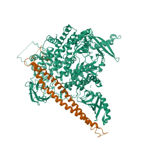
- PIK3CA encodes the p110-alpha catalytic subunit of PI3K, a lipid kinase that phosphorylates phosphatidylinositol 4,5-bisphosphate (PtdIns(4,5)P2) to generate phosphatidylinositol 3,4,5-trisphosphate (PIP3), a critical second messenger.
Related references: PMID:15135396, PMID:23936502, PMID:28676499 - PIP3 recruits pleckstrin homology (PH) domain-containing proteins like AKT1 and PDPK1 to the membrane, initiating signaling cascades that promote cell growth, proliferation, survival, motility, and cytoskeletal reorganization.
- PIK3CA displays intrinsic serine-protein kinase activity, enabling autophosphorylation of the regulatory p85-alpha subunit and phosphorylation of substrates such as 4EBP1 and H-Ras, potentially modulating downstream pathways.
Related references: PMID:23936502, PMID:28676499 - It is activated by receptor tyrosine kinases (e.g., EGFR, insulin receptor, VEGFR) and RAS, playing essential roles in growth factor signaling, angiogenesis, and vascular development.
- PIK3CA is classified as a proto-oncogene; somatic gain-of-function mutations are frequently detected in a wide range of human cancers, driving uncontrolled PI3K/AKT pathway activation.
- The protein is also implicated in phagocytosis and pinocytosis, although the exact mechanisms remain partially characterized (By similarity).
Experimental Guidance and Technical Tips
- The immunogen comprises amino acids 401–600 of human PIK3CA; this central region is well-conserved across human, mouse, and rat, consistent with the reported cross-reactivity.
- For Western blot, PIK3CA migrates at a predicted molecular weight of ~124 kDa; ensure sufficient gel electrophoresis resolution and transfer.
- Validate antibody performance in relevant positive control lysates (e.g., cancer cell lines overexpressing PIK3CA or stimulated with growth factors) before use in experimental samples.
- For immunohistochemistry (IHC-P), optimize antigen retrieval protocols (e.g., heat-induced epitope retrieval) and consider titrating antibody concentration to achieve specific staining.
- Because PIK3CA is involved in numerous signaling networks, include appropriate pathway inhibitors in functional studies if using the antibody for co-detection or pull-down.
CamelBio: Your One-Stop Sourcing Bridge
CamelBio supports diagnostic manufacturers and researchers with a broad portfolio of IVD raw materials, including validated antibody pairs, monoclonal/polyclonal antibodies, and ancillary reagents for the PI3K/AKT pathway. This PIK3CA rabbit polyclonal antibody is part of CamelBio’s commitment to providing high-quality tools for cancer research, cell signaling, and assay development. From target discovery to clinical translation, CamelBio delivers integrated sourcing solutions that streamline your workflow.
Product Datasheet
Anti-PIK3CA Polyclonal Antibody for WB, IHC-P, IF/ICC, ELISA - P42336
REQUEST A QUOTE
Our professional team will reply to you within one business day. Please feel free to contact us!