Phosphorylated Antibodies
Anti-Phospho-ZAP70 (Y493) Rabbit Polyclonal Antibody for WB, IHC-P, IF/ICC, ELISA - P43403
Item Number : CM0001417
Price varies based on specs and customizations
- Application
- WB, IHC-P, IF/ICC, ELISA
- Cross Reactivity
- Human, Mouse, Rat
- Protein Weight
- 70kDa

Shipping:
Contact us to get shipping details Enjoy On-time Dispatch Guarantee.
Why Choose Us
Easy ordering process, quality products, and dedicated support for your business success.
Core Product Specifications and Parameters
| Parameter | Value |
|---|---|
| Product Name | Phospho-ZAP70-Y493 Rabbit pAb |
| Remarks/Alias | SRK; STD; TZK; STCD; IMD48; ADMIO2; ZAP-70; Phospho-ZAP70-Y493 |
| Species | Human |
| Gene ID (Human) | 7535 |
| Gene ID | 7535 |
| Immunogen | Synthetic peptide: A synthetic phosphorylated peptide around Y493 of human ZAP70 (NP_001070.2). |
| Source | Rabbit |
| Category | Phosphorylated Antibodies |
| Application | WB, IHC-P, IF/ICC, ELISA |
| Cross Reactivity | Human, Mouse, Rat |
| SWISS | P43403 |
| Protein Weight | 70kDa |
| Shipping | Ice bag |
Biological Background: ZAP70 Function and Localization
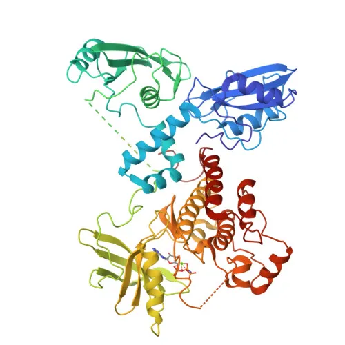
- ZAP70 (70 kDa zeta-chain associated protein) is a cytoplasmic tyrosine kinase essential for adaptive immune regulation, mediating T-cell receptor (TCR) signaling upon antigen presentation (PubMed:7509083). Related references: PMID:7509083
- The protein contains tandem SH2 domains that bind phosphorylated ITAM motifs on CD3 chains, relieving autoinhibition and enabling LCK-mediated phosphorylation at Tyr493 for full activation.
- Upon activation, ZAP70 phosphorylates key adaptors LAT and LCP2, triggering downstream cascades that regulate T-cell proliferation, cytokine production, and cytoskeletal reorganization.
- ZAP70 is involved in thymocyte development, promoting survival and cell-cycle progression of CD4/CD8 double-negative cells before positive selection.
- It also contributes to B-cell development and activation through B-cell receptor signaling, highlighting broader lymphoid roles.
- Subcellular localization is primarily cytoplasmic, with recruitment to the plasma membrane upon TCR engagement; it also participates in TCR internalization via CBL-mediated ubiquitination.
- Expressed predominantly in T-cells, natural killer cells, early thymocytes, and pro/pre B-cells, with genetic defects causing severe combined immunodeficiency (SCID) and autoimmune conditions.
- Post-translational modifications include tyrosine phosphorylation, acetylation, and ubiquitination, critical for its dynamic regulation.
Experimental Guidance and Technical Tips
- As a phospho-specific antibody targeting Tyr493, ensure samples are treated with phosphatase inhibitors during lysis and consider phosphatase treatment as a negative control.
- For Western blot, the predicted molecular weight is approximately 70 kDa; use an appropriate gel percentage (e.g., 8-10%) for optimal resolution.
- Validate antibody specificity in your model system using peptide competition or genetic knockdown/knockout approaches.
- For immunohistochemistry (IHC-P), antigen retrieval may be required; test pH 6 or pH 9 retrieval buffers.
- In immunofluorescence (IF/ICC), monitor membrane localization upon stimulation; permeabilization and fixation conditions should be optimized.
- ELISA and other applications may require titration to determine the optimal working concentration.
CamelBio: Your One-Stop Sourcing Bridge
CamelBio is a dedicated one-stop sourcing partner for IVD raw materials, supporting diagnostic and research communities from concept to clinic. This anti-phospho-ZAP70 (Tyr493) antibody, targeting a key node in adaptive immunity and T-cell signaling, exemplifies our capability to supply validated phospho-specific antibodies for demanding applications. We also offer comprehensive antibody development services, bulk reagent supply, and rare target sourcing to streamline your project workflows.
REQUEST A QUOTE
Our professional team will reply to you within one business day. Please feel free to contact us!