Phosphorylated Antibodies
Anti-Phospho-TAK1-S439 Rabbit Monoclonal Antibody for WB - O43318
Item Number : CM0000553
Price varies based on specs and customizations
- Application
- WB, ELISA
- Cross Reactivity
- Mouse
- Protein Weight
- 67kDa

Shipping:
Contact us to get shipping details Enjoy On-time Dispatch Guarantee.
Why Choose Us
Easy ordering process, quality products, and dedicated support for your business success.
Core Product Specifications and Parameters
| Core Parameter | Value |
|---|---|
| Product Name | Phospho-TAK1-S439 Rabbit mAb |
| Remarks/Alias | CSCF; FMD2; TAK1; MEKK7; TGF1a; Phospho-TAK1-S439 |
| Species | Human |
| GeneID (Human) | 6885 |
| GeneID | 6885 |
| Immunogen | A synthetic phosphorylated peptide around S439 of human TAK1 (O43318) |
| Source | Rabbit |
| Category | Phosphorylated Antibodies |
| Application | WB, ELISA |
| Cross Reactivity | Mouse |
| SWISS | O43318 |
| Protein Weight | 67kDa |
| Shipping | Ice bag |
Biological Background: TAK1 Function and Localization
- Mitogen-activated protein kinase kinase kinase 7 (MAP3K7), also known as TAK1, is an essential serine/threonine kinase in the MAP kinase signal transduction pathway, mediating cellular responses to environmental changes.
Related references: PMID:10094049 | PMID:11460167 | PMID:12589052 - TAK1 integrates signals from diverse stimuli, including TRAF6, IL-1, TGFB, BMP2/4, TLRs, CD40, and BCR, and phosphorylates downstream MAP2Ks (MEK1, MKK3, MKK6, MKK7) to activate p38 MAPK and JNK pathways, controlling AP-1 transcription.
Related references: PMID:11460167 | PMID:8663074 | PMID:16893890 | PMID:9079627 - Independently of MAP2Ks, TAK1 activates NF-κB by recruiting TAB2/TAB3 to polyubiquitin chains, leading to phosphorylation and activation of the IKK complex.
Related references: PMID:12589052 | PMID:8663074 | PMID:10094049 | PMID:11460167 - Under osmotic stress, TAK1 specifically activates MAPK8/JNK1 but not NF-κB.
Related references: PMID:16893890 - TAK1 localizes to the cytoplasm and cell membrane, consistent with its role in receptor-proximal signaling.
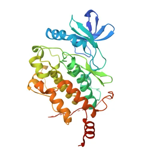
- Phosphorylation of TAK1, particularly at Ser439, is critical for its activation; this antibody specifically recognizes the phospho-S439 form. (Keywords: Phosphoprotein, Serine/threonine-protein kinase)
- TAK1 also promotes TRIM5-mediated retrovirus restriction, regulates RIPK1-dependent necroptosis/apoptosis, and phosphorylates STING1 for antiviral signaling.
Related references: PMID:21512573 | PMID:37832545
Experimental Guidance and Technical Tips
- The immunogen is a synthetic phosphopeptide around S439; ensure phospho-specificity by including non-phosphorylated peptide controls or phosphatase-treated samples in your assays.
- For Western blot, consider using a loading control and validate in cell lines stimulated with cytokines (e.g., IL-1β, TGF-β) or stress inducers to confirm detection of the phosphorylated band at ~67 kDa.
- For ELISA, peptide competition assays can help verify that the signal is specifically due to the phospho-epitope.
- Include phosphatase inhibitors during sample preparation to preserve the phosphorylation signal.
- Cross-reactivity with mouse TAK1 allows translational studies in mouse models; validate expected phosphorylation patterns in mouse tissues.
CamelBio: Your One-Stop Sourcing Bridge
CamelBio provides diagnostic manufacturers and research laboratories with one-stop access to premium IVD raw materials, including validated antibody pairs, optimized monoclonal antibodies, and bulk ancillary reagents. For TAK1 signaling studies, we offer this phospho-specific rabbit mAb and can source additional key reagents to support your workflow. As your procurement partner, we help streamline supply from concept to clinic, ensuring consistent quality and reliable access to rare targets and custom raw materials.
REQUEST A QUOTE
Our professional team will reply to you within one business day. Please feel free to contact us!