Phosphorylated Antibodies
Anti-Phospho-STK3/MST2-T384 Rabbit Polyclonal Antibody for WB, ELISA - Q13188
Item Number : CM0001335
Price varies based on specs and customizations
- Application
- WB, ELISA
- Cross Reactivity
- Human, Mouse, Rat
- Protein Weight
- 56kDa

Shipping:
Contact us to get shipping details Enjoy On-time Dispatch Guarantee.
Why Choose Us
Easy ordering process, quality products, and dedicated support for your business success.
Core Product Specifications and Parameters
| Parameter | Value |
|---|---|
| Product Name | Phospho-STK3/MST2-T384 Rabbit pAb |
| Remarks/Alias | KRS1; MST2; Phospho-STK3/MST2-T384 |
| Species | Human |
| Gene ID (Human) | 6788 |
| Gene ID | 6788 |
| Immunogen | Synthetic Peptide: A synthetic phosphorylated peptide around T384 of human STK3/MST2 (NP_006272.2) |
| Source | Rabbit |
| Category | Phosphorylated Antibodies |
| Application | WB, ELISA |
| Cross Reactivity | Human, Mouse, Rat |
| SWISS | Q13188 |
| Protein Weight | 56kDa |
| Shipping | Ice bag |
Biological Background: Serine/threonine-protein kinase 3 (STK3) Function and Localization
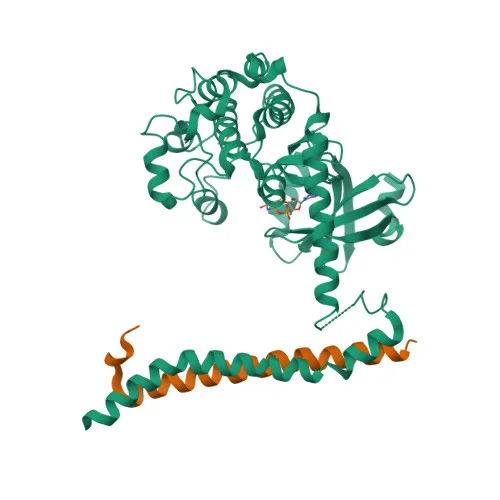
- STK3 (also known as MST2 or Krs-1) is a serine/threonine-protein kinase that acts as a pro-apoptotic stress-activated kinase. It cleaves and translocates to the nucleus to induce chromatin condensation and DNA fragmentation. Related references: PMID:11278283, PMID:8566796, PMID:8816758
- As a core component of the Hippo signaling pathway, STK3 phosphorylates and activates LATS1/2, which in turn phosphorylate and inactivate YAP1 and WWTR1/TAZ, regulating cell proliferation, apoptosis, and organ size. Related references: PMID:15688006, PMID:16930133, PMID:23972470
- STK3/MST2 and its homolog STK4/MST1 repress proliferation of mature hepatocytes and prevent activation of facultative adult liver stem cells, thus inhibiting tumor formation.
- Phosphorylates NEK2 and plays a role in centrosome disjunction by regulating NEK2 localization and its ability to phosphorylate CROCC and CEP250. Related references: PMID:21076410, PMID:21723128
- Positively regulates RAF1 activation by suppressing inhibitory phosphorylation of RAF1 on Ser-259. Related references: PMID:20212043
- Phosphorylates MOBKL1A and RASSF2 and cooperates with MOBKL1B to activate STK38. Related references: PMID:19525978, PMID:18328708, PMID:18362890
- Predominantly localized in the cytoplasm and nucleus, and also associates with the centrosome. Expressed at high levels in kidney, skeletal muscle, and placenta tissues.
Experimental Guidance and Technical Tips
- The immunogen is a synthetic phosphorylated peptide corresponding to residues around Thr384 of human STK3. For Western blot, consider using a phospho-specific blocking and detection protocol to preserve epitope recognition; pre-absorption with the non-phosphorylated peptide may help confirm signal specificity.
- Cross-reactivity with mouse and rat STK3 is indicated; validate the antibody in your specific model system, as sequence homology around Thr384 is high.
- For ELISA, a phospho-peptide–specific immobilization strategy may improve detection; ensure coating conditions do not dephosphorylate the peptide.
- Standard anti-rabbit secondary antibodies are suitable for detection; optimize antibody dilutions for your system.
CamelBio: Your One-Stop Sourcing Bridge
CamelBio serves as a comprehensive sourcing partner for diagnostic manufacturers and research labs, offering validated antibody pairs, monoclonal and polyclonal antibodies, bulk ancillary reagents, and custom raw‑material procurement. This phospho-specific STK3 antibody supports research into apoptosis and Hippo pathway signaling, and we facilitate the complete supply chain from reagent selection to assay development.
REQUEST A QUOTE
Our professional team will reply to you within one business day. Please feel free to contact us!