Phosphorylated Antibodies
Anti-Phospho-RIPK1 (S166) Rabbit Polyclonal Antibody for WB and ELISA - Q13546
Item Number : CM0001155
Price varies based on specs and customizations
- Application
- WB, ELISA
- Cross Reactivity
- Human, Mouse
- Protein Weight
- 76 kDa

Shipping:
Contact us to get shipping details Enjoy On-time Dispatch Guarantee.
Why Choose Us
Easy ordering process, quality products, and dedicated support for your business success.
Core Product Specifications and Parameters
| Parameter | Value |
|---|---|
| Product Name | Phospho-RIPK1/RIP-S166 Rabbit pAb |
| Remarks/Alias | RIP; RIP1; AIEFL; IMD57; RIP-1; Phospho-RIPK1/RIP-S166 |
| Species | Human |
| Gene ID | 8737 |
| Immunogen | A synthetic phosphorylated peptide around S166 of human RIPK1/RIP (NP_003795.2) |
| Source | Rabbit |
| Category | Phosphorylated Antibodies |
| Application | WB, ELISA |
| Cross Reactivity | Human, Mouse |
| SWISS | Q13546 |
| Protein Weight | 76 kDa |
| Shipping | Ice bag |
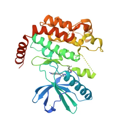
Biological Background: RIPK1 Function and Localization
- RIPK1 is a serine-threonine kinase that acts as a key regulator of TNF-mediated apoptosis, necroptosis, and inflammatory pathways [PMID:17703191, PMID:24144979, PMID:31827280]. Related references: PMID:17703191 PMID:24144979 PMID:31827280
- The kinase activity of RIPK1 is essential for regulating cell death, including the assembly of apoptotic and necroptotic complexes [PMID:11101870, PMID:19524512, PMID:19524513]. Related references: PMID:11101870 PMID:19524512 PMID:19524513
- RIPK1 also has kinase-independent scaffold functions, promoting cell survival by activating NF-kappa-B when recruited to the TNF-R1 signaling complex.
- During embryonic development, RIPK1 is required to inhibit apoptosis and necroptosis, preventing aberrant cell death.
- The protein localizes to both the cytoplasm and the cell membrane.
- RIPK1 undergoes multiple post-translational modifications, including phosphorylation, ubiquitination, and proteolytic cleavage, which dictate its pro-survival or pro-death functions.
- RIPK1 is involved in the inflammatory response, promoting production of pro-inflammatory cytokines such as IL-6 [PMID:31827280, PMID:31827281]. Related references: PMID:31827280 PMID:31827281
Experimental Guidance and Technical Tips
- The immunogen is a synthetic phosphopeptide around Ser166 of human RIPK1; the antibody is expected to specifically recognize RIPK1 phosphorylated at this site.
- For Western blot, induce RIPK1 phosphorylation using TNF or other stimuli in your experimental system prior to lysis. Consider using phosphatase inhibitors in lysis buffer.
- Validate specificity by comparing signals from phosphatase-treated samples or by peptide competition with the non-phosphorylated control peptide.
- For ELISA, the phospho-peptide can be used as coating antigen; confirm signal reduction upon dephosphorylation.
- Cross-reactivity with mouse is indicated; verify detection in mouse tissues or cells, and optimize antibody dilution for each species.
CamelBio: Your One-Stop Sourcing Bridge
CamelBio is your one-stop IVD raw-material provider, supplying validated antibody pairs, optimized monoclonal and polyclonal antibodies, and ancillary reagents to diagnostic manufacturers and research labs. Our expertise in cell death and inflammatory signaling supports your RIPK1-related assay development, from early R&D to clinical validation. Whether you need bulk quantities or rare-target sourcing, CamelBio streamlines your path from concept to clinic.
REQUEST A QUOTE
Our professional team will reply to you within one business day. Please feel free to contact us!