Phosphorylated Antibodies
Anti-Phospho-RIPK1 (S166) Rabbit Monoclonal Antibody for WB, ELISA - Q13546
Item Number : CM0001043
Price varies based on specs and customizations
- Application
- WB, ELISA
- Cross Reactivity
- Human
- Protein Weight
- 75kDa

Shipping:
Contact us to get shipping details Enjoy On-time Dispatch Guarantee.
Why Choose Us
Easy ordering process, quality products, and dedicated support for your business success.
Core Product Specifications and Parameters
| Parameter | Value |
|---|---|
| Product Name | Phospho-RIPK1-S166 Rabbit mAb |
| Remarks/Alias | RIP; RIP1; AIEFL; IMD57; RIP-1; Phospho-RIPK1-S166 |
| Species | Human |
| Gene ID (Human) | 8737 |
| Gene ID | 8737 |
| Immunogen | Synthetic peptide; A synthetic phosphorylated peptide around S166 of human RIPK1 (NP_003795.2) |
| Source | Rabbit |
| Category | Phosphorylated Antibodies |
| Application | WB, ELISA |
| Cross Reactivity | Human |
| SWISS | Q13546 |
| Protein Weight | 75kDa |
| Shipping | Ice bag |
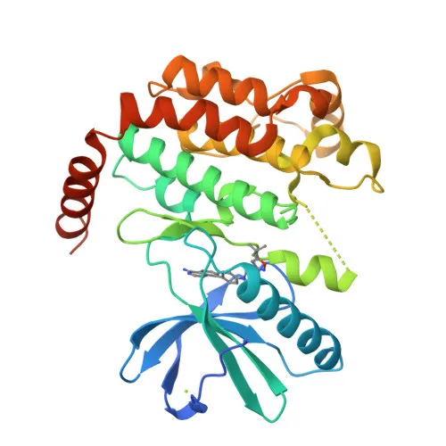
Biological Background: Receptor-interacting serine/threonine-protein kinase 1 Function and Localization
- RIPK1, also known as RIP, RIP-1, or cell death protein RIP, is a serine-threonine kinase encoded by the RIPK1 gene; alternative splicing produces multiple protein isoforms.
- It serves as a key regulator of TNF-mediated apoptosis, necroptosis, and inflammatory pathways. Related references: PMID:17703191, PMID:24144979, PMID:31827280.
- RIPK1 exhibits kinase activity-dependent functions controlling cell death and kinase-independent scaffold functions that promote inflammatory signaling and cell survival. Related references: PMID:11101870, PMID:19524512, PMID:19524513.
- Upon TNF stimulation, RIPK1 is recruited to the TNF-R1 signaling complex (complex I) where it acts as a scaffold to activate canonical NF-κB and promote cell survival.
- The kinase activity of RIPK1 drives the assembly of death-inducing complexes: complex IIa (RIPK1-FADD-CASP8) for apoptosis and complex IIb (RIPK1-RIPK3-MLKL) for necroptosis.
- RIPK1 phosphorylates RIPK3, leading to reciprocal auto- and trans-phosphorylation. Related references: PMID:19524513.
- It phosphorylates DAB2IP at Ser-728, thereby activating the MAP3K5-JNK apoptotic cascade. Related references: PMID:15310755, PMID:17389591.
- Subcellularly, RIPK1 is found in the cytoplasm and associated with the cell membrane; it undergoes phosphorylation, ubiquitination, and other post-translational modifications that regulate its signaling functions.
Experimental Guidance and Technical Tips
- The immunogen is a synthetic phosphorylated peptide corresponding to residues around serine 166 of human RIPK1; thus, the antibody specifically recognizes RIPK1 when phosphorylated at this site.
- For Western blotting, use lysates from human cell lines or tissues, as the antibody is reported to react with human RIPK1; consider including phosphatase inhibitors during sample preparation to preserve phosphorylation.
- ELISA applications may require optimization of coating conditions and detection of phosphorylated RIPK1 in cell lysates or recombinant protein; validate with positive and negative controls for phosphorylation.
- The predicted molecular weight of RIPK1 is ~75 kDa; however, post-translational modifications may cause slight shifts in migration.
CamelBio: Your One-Stop Sourcing Bridge
CamelBio offers a comprehensive portfolio of IVD raw materials, supporting diagnostic manufacturers and researchers in the cell death and inflammatory disease areas. Our catalog includes validated phosphorylation-specific antibodies, recombinant proteins, and ancillary reagents. For specialized targets like phospho-RIPK1, CamelBio facilitates sourcing of rare raw materials and bulk supplies, ensuring seamless integration into your assay development pipeline.
REQUEST A QUOTE
Our professional team will reply to you within one business day. Please feel free to contact us!