Phosphorylated Antibodies
Anti-Phospho-PTEN-S380 Rabbit Monoclonal Antibody for WB, ELISA - P60484
Item Number : CM0000956
Price varies based on specs and customizations
- Application
- WB, ELISA
- Cross Reactivity
- Mouse
- Protein Weight
- 47kDa

Shipping:
Contact us to get shipping details Enjoy On-time Dispatch Guarantee.
Why Choose Us
Easy ordering process, quality products, and dedicated support for your business success.
Core Product Specifications and Parameters
| Parameter | Value |
|---|---|
| Product Name | Phospho-PTEN-S380 Rabbit mAb |
| Remarks/Alias | BZS; DEC; CWS1; GLM2; MHAM; TEP1; MMAC1; PTEN1; 10q23del; PTENbeta; Phospho-PTEN-S380 |
| Species | Human |
| Gene ID | 5728 |
| Immunogen | Synthetic Peptide: A synthetic phosphorylated peptide around S380 of human PTEN (P60484) |
| Source | Rabbit |
| Category | Phosphorylated Antibodies |
| Application | WB, ELISA |
| Cross Reactivity | Mouse |
| SWISS | P60484 |
| Protein Weight | 47kDa |
| Shipping | Ice bag |
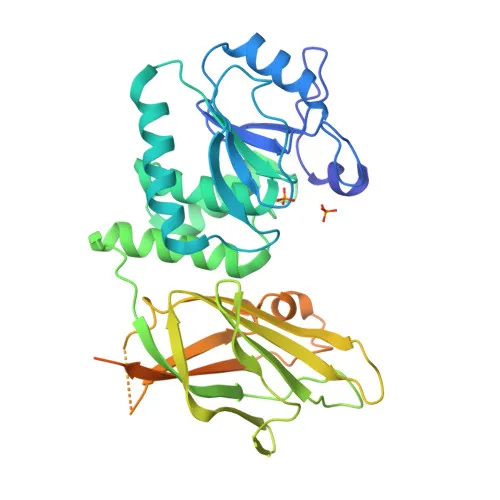
Biological Background: PTEN Function and Localization
- PTEN (phosphatase and tensin homolog) is a dual-specificity phosphatase that functions as both a lipid and protein phosphatase, and is a well-known tumor suppressor.
- As a lipid phosphatase, it dephosphorylates phosphatidylinositol 3,4,5-trisphosphate (PIP3) to generate PIP2, thereby directly antagonizing the PI3K-AKT signaling pathway and regulating cell survival, proliferation, and metabolism. Related references: PMID:16824732 PMID:26504226 PMID:9593664
- Also exhibits protein phosphatase activity, dephosphorylating tyrosine-, serine-, and threonine-phosphorylated proteins, including focal adhesion kinase, which influences cell migration and adhesion. Related references: PMID:22279049
- PTEN is phosphorylated at multiple residues, including S380, which regulates its activity, conformation, and subcellular localization; phosphorylation at the C-terminal tail reduces its membrane binding and may modulate tumor suppressor function.
- Subcellular localization is dynamic: present in the cytoplasm, nucleus, PML bodies, and at the postsynaptic density in neurons; nuclear monoubiquitinated PTEN exhibits enhanced apoptotic potential. Related references: PMID:10468583 PMID:18716620
- Widely expressed in adult tissues including heart, brain, placenta, lung, liver, muscle, kidney, and pancreas, underscoring its fundamental roles in cellular homeostasis.
- A key modulator of AKT-mTOR signaling during neurogenesis and synaptic plasticity, and required for long-term depression (LTD) in hippocampal neurons.
- Genetic alterations or dysregulation of PTEN are linked to multiple cancers and developmental disorders, highlighting its clinical significance as a therapeutic target.
Experimental Guidance and Technical Tips
- For Western blot, use the antibody at a recommended starting dilution (e.g., 1:500-1:2000) after validation in the experimental system; include phosphatase inhibitors during sample preparation to preserve phosphorylation states.
- In ELISA, confirm optimal coating antigen concentration and antibody dilution through checkerboard titration.
- Validate cross-reactivity with mouse PTEN in appropriate tissue or cell lysates; confirm specificity via peptide competition or treatment with phosphatases.
- Monitor multiple phosphorylation sites to study PTEN regulation interplay; S380 phosphorylation may impact its protein interactions and function.
CamelBio: Your One-Stop Sourcing Bridge
CamelBio provides diagnostic manufacturers, labs, and research institutes with one-stop access to IVD raw materials, technical services, and consulting—covering every stage from concept to clinic. Our portfolio includes validated antibody pairs, optimized monoclonal/polyclonal antibodies, bulk ancillary reagents, and rare target raw-material sourcing. For PTEN signaling studies, we offer a reliable supply of high-quality phospho-specific and pan antibodies, supporting your research and diagnostic development needs.
Product Datasheet
Anti-Phospho-PTEN-S380 Rabbit Monoclonal Antibody for WB, ELISA - P60484
REQUEST A QUOTE
Our professional team will reply to you within one business day. Please feel free to contact us!