Phosphorylated Antibodies
Anti-Phospho-PAK1/2/3 (S144/S141/S139) Rabbit Monoclonal Antibody for WB and ELISA - Q13153 / Q13177 / O75914
Item Number : CM0001673
Price varies based on specs and customizations
- Application
- WB, ELISA
- Cross Reactivity
- Human, Mouse, Rat
- Protein Weight
- 65kDa

Shipping:
Contact us to get shipping details Enjoy On-time Dispatch Guarantee.
Why Choose Us
Easy ordering process, quality products, and dedicated support for your business success.
Core Product Specifications and Parameters
| Parameter | Value |
|---|---|
| Product Name | Phospho-PAK1/2/3-S144/S141/S139 Rabbit mAb |
| Remarks/Alias | PAKalpha; Phospho-PAK1/2/3-S144/S141/S139 |
| Species | Human |
| GeneID (Human) | 5058/5062/5063 |
| GeneID | 5058/5062/5063 |
| Immunogen | A synthetic phosphorylated peptide around S144 of human PAK1 (Q13153) |
| Source | Rabbit |
| Category | Phosphorylated Antibodies |
| Application | WB, ELISA |
| Cross Reactivity | Human, Mouse, Rat |
| SWISS | Q13153/Q13177/O75914 |
| Protein Weight | 65kDa |
| Shipping | Ice bag |
Biological Background: PAK1 Function and Localization
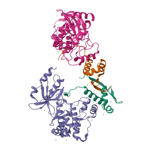
- PAK1 (p21-activated kinase 1, alpha-PAK) is a serine/threonine-protein kinase that, together with PAK2 and PAK3, belongs to the PAK family. This antibody recognizes the phosphorylated form of the conserved serine residue equivalent to S144 in PAK1, S141 in PAK2, and S139 in PAK3, which are critical for kinase activation.
- Activated by the Rho GTPases CDC42 and RAC1, PAK1 links extracellular signals to intracellular pathways controlling cytoskeletal dynamics, cell adhesion, and migration.
Related references: PMID:8805275 PMID:9528787 - PAK1 reorganizes the actin cytoskeleton by promoting stress fiber and focal adhesion formation, thereby regulating cell shape and motility.
Related references: PMID:9032240 PMID:9395435 - The kinase regulates apoptosis through RAF1 phosphorylation, which in turn phosphorylates BAD and promotes its mitochondrial sequestration.
Related references: PMID:11733498 - Subcellularly, PAK1 localizes to the cytoplasm, cell junctions, lamellipodia, ruffle membranes, invadopodia, nucleus, and centrosome, matching its multifaceted roles in signaling and cell structure.
- Post-translational modifications include autophosphorylation at multiple serine/threonine residues (including S144) and acetylation, which modulate activity and localization.
- Clinically, PAK1 is overexpressed in gastric cancer, promoting invasion and migration, and has been linked to neurological disorders.
Related references: PMID:25766321
Experimental Guidance and Technical Tips
- For Western blotting, use protease and phosphatase inhibitor cocktails during cell/tissue lysis to preserve phosphorylation status. The expected band for PAK1 is approximately 65 kDa, but slight size variations may occur across species or due to post‑translational modifications.
- Specificity of phospho‑signal can be confirmed by treating samples with λ‑phosphatase or by peptide competition assays using the non‑phosphorylated immunogen peptide.
- In ELISA, use a phosphorylated peptide corresponding to the immunogen as the coating antigen, and include appropriate controls (non‑phosphorylated peptide, blank) to assess antibody specificity.
- Since the antibody recognizes a conserved phospho‑site shared by PAK1/2/3, it is advisable to verify isoform identity in your system using isoform‑specific total‑PAK antibodies or siRNA knockdown.
CamelBio: Your One-Stop Sourcing Bridge
CamelBio streamlines IVD raw-material procurement with a comprehensive portfolio of validated phospho‑specific antibodies, including this anti‑PAK1/2/3 (S144/S141/S139) monoclonal antibody, tailored for intracellular signaling research. Beyond single reagents, we offer matched antibody pairs, bulk ancillary reagents, and custom sourcing for challenging targets—connecting diagnostic manufacturers with critical components from discovery to commercialization.
REQUEST A QUOTE
Our professional team will reply to you within one business day. Please feel free to contact us!