Phosphorylated Antibodies
Anti-Phospho-MST1/STK4-T387 Rabbit Polyclonal Antibody for Western Blot and ELISA - Q13043
Item Number : CM0001244
Price varies based on specs and customizations
- Application
- WB, ELISA
- Cross Reactivity
- Human, Mouse, Rat
- Protein Weight
- 56kDa

Shipping:
Contact us to get shipping details Enjoy On-time Dispatch Guarantee.
Why Choose Us
Easy ordering process, quality products, and dedicated support for your business success.
Core Product Specifications and Parameters
| Parameter | Value |
|---|---|
| Product Name | Phospho-MST1/STK4-T387 Rabbit pAb |
| Remarks/Alias | KRS2; MST1; YSK3; Phospho-MST1/STK4-T387 |
| Species | Human |
| Gene ID (Human) | 6789 |
| Immunogen | A synthetic phosphorylated peptide around T387 of human MST1/STK4 (NP_006273.1) |
| Source | Rabbit |
| Category | Phosphorylated Antibodies |
| Application | WB, ELISA |
| Cross Reactivity | Human, Mouse, Rat |
| SWISS | Q13043 |
| Protein Weight | 56kDa |
| Shipping | Ice bag |
Biological Background: MST1/STK4 Function and Localization
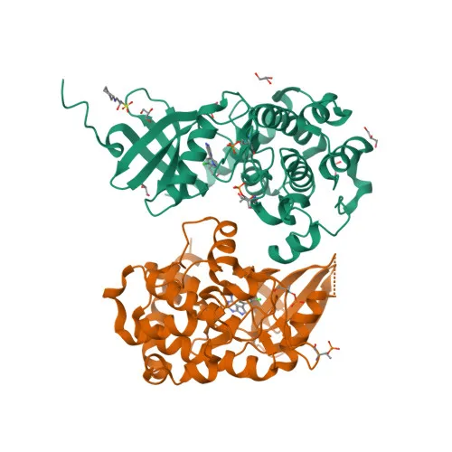
- Serine/threonine-protein kinase 4 (STK4), also known as MST-1, Krs-2, and YSK3, is encoded by the STK4 gene and belongs to the STE20 kinase family.
- Acts as a stress-activated, pro-apoptotic kinase that, following caspase cleavage, translocates to the nucleus to induce chromatin condensation and internucleosomal DNA fragmentation.
- Forms the core of the Hippo signaling pathway by complexing with SAV1 to phosphorylate and activate LATS1/2, which in turn phosphorylate YAP1/WWTR1 to restrict proliferation and promote apoptosis.
- Phosphorylates diverse substrates including histone H2B (Ser-14), FOXO3, FOXO1, SIRT1, and AR; also acts as an inhibitor of PKB/AKT1.
- Localizes primarily to the cytoplasm but enters the nucleus upon receipt of apoptotic signals.
- Ubiquitously expressed, with elevated protein levels observed in prostate cancer from normal to malignant states.
- Post-translational modifications include acetylation and phosphorylation; alternative splicing produces multiple isoforms.
Experimental Guidance and Technical Tips
- Confirm phospho-specificity by using a non-phosphorylated peptide blocking control or phosphatase treatment in western blot experiments.
- Expected band size in western blot is approximately 56 kDa; include positive controls (e.g., apoptotic cells) and relevant negative controls.
- For ELISA, consider coating with the phospho-peptide immunogen and a non-phospho peptide to assess specificity and titer.
- Validate the antibody in your target species (human, mouse, rat) and tissue types under appropriate conditions.
CamelBio: Your One-Stop Sourcing Bridge
CamelBio provides one-stop access to validated IVD raw materials, including phospho-specific antibodies like anti-Phospho-MST1/STK4-T387 pAb, supporting research into apoptosis and the Hippo signaling pathway. We offer bulk sourcing of optimized monoclonal and polyclonal antibodies, matched antibody pairs, and ancillary reagents, and can assist with rare target raw-material procurement to accelerate your diagnostic development.
REQUEST A QUOTE
Our professional team will reply to you within one business day. Please feel free to contact us!