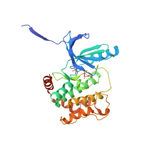
Phosphorylated Antibodies
Anti-Phospho-MAP2K4 (S257/T261) Rabbit Polyclonal Antibody for WB and ELISA - P45985
Item Number : CM0001436
Price varies based on specs and customizations
- Application
- WB, ELISA
- Cross Reactivity
- Human, Mouse, Rat
- Protein Weight
- 44kDa

Shipping:
Contact us to get shipping details Enjoy On-time Dispatch Guarantee.
Why Choose Us
Easy ordering process, quality products, and dedicated support for your business success.
Core Product Specifications and Parameters
| Parameter | Value |
|---|---|
| Product Name | Phospho-MAP2K4-S257/T261 Rabbit pAb |
| Remarks/Alias | JNKK; MEK4; MKK4; SEK1; SKK1; JNKK1; SERK1; MAPKK4; PRKMK4; SAPKK1; SAPKK-1; Phospho-MAP2K4-S257/T261 |
| Species | Human |
| Gene ID | 6416 |
| Immunogen | Synthetic peptide (A synthetic phosphorylated peptide around S257 & T261 of human MAP2K4 (NP_003001.1)) |
| Source | Rabbit |
| Category | Phosphorylated Antibodies |
| Application | WB, ELISA |
| Cross Reactivity | Human, Mouse, Rat |
| SWISS | P45985 |
| Protein Weight | 44kDa |
| Shipping | Ice bag |
Biological Background: MAP2K4 Function and Localization
- MAP2K4 (Dual specificity mitogen-activated protein kinase kinase 4), also known as MEK4, MKK4, SEK1, JNKK1, or SAPKK1, is a Ser/Thr and Tyr dual-specificity protein kinase.
- It is an essential component of the stress-activated protein kinase/c-Jun N-terminal kinase (SAP/JNK) signaling pathway, directly activating MAPK8/JNK1, MAPK9/JNK2, and MAPK10/JNK3.
- MAP2K4 preferentially phosphorylates the Tyr residue of the Thr-Pro-Tyr motif in JNKs, while MAP2K7/MKK7 phosphorylates the Thr residue; both kinases synergize for full JNK activation.
- Beyond JNKs, MAP2K4 also activates p38 MAPKs (MAPK11, MAPK12, MAPK13, MAPK14), playing a broader role in stress and inflammatory responses.
- The kinase is required for maintaining peripheral lymphoid homeostasis and is involved in mitochondrial death signaling, including cytochrome c release and apoptosis.
- Subcellular localization includes both the cytoplasm and the nucleus.
- It is widely expressed, with particularly abundant expression in skeletal muscle.
- Post-translational modifications include phosphorylation (at Ser257, Thr261, and other sites), acetylation, and methylation, regulating its activity and interactions.
Experimental Guidance and Technical Tips
- The immunogen is a synthetic phosphorylated peptide encompassing S257 and T261 of human MAP2K4, making this antibody suitable for detecting the dual-phosphorylated, active form of the kinase.
- For Western blot, consider using phosphatase‑treated lysates or non‑phospho peptide competition to confirm phospho‑specificity. A loading control is recommended for quantitative analyses.
- For ELISA, validate the antibody in the relevant sample system with appropriate blocking and wash buffers to minimize non‑specific binding.
- The antibody is reported to cross‑react with mouse and rat orthologs, which share high sequence identity in the immunogen region; verify in your specific model system.
- Phosphorylation at S257 and T261 correlates with MAP2K4 activation, so this antibody can be a valuable tool for monitoring stress‑induced signaling events.
CamelBio: Your One-Stop Sourcing Bridge
CamelBio serves as a one‑stop IVD raw‑material partner, providing diagnostic manufacturers with validated antibody pairs, optimized monoclonal and polyclonal antibodies, and bulk ancillary reagents. This phospho‑MAP2K4 antibody is part of our extensive catalog supporting stress signaling and apoptosis research, and our sourcing team is ready to assist with custom raw‑material needs to accelerate your assay development.
REQUEST A QUOTE
Our professional team will reply to you within one business day. Please feel free to contact us!