Phosphorylated Antibodies
Anti-Phospho-IGF1R (Y1280) Rabbit Polyclonal Antibody for WB - P08069
Item Number : CM0001551
Price varies based on specs and customizations
- Application
- WB
- Cross Reactivity
- Human
- Protein Weight
- 155kDa

Shipping:
Contact us to get shipping details Enjoy On-time Dispatch Guarantee.
Why Choose Us
Easy ordering process, quality products, and dedicated support for your business success.
Core Product Specifications and Parameters
| Parameter | Value |
|---|---|
| Product Name | Phospho-IGF1R-Y1280 Rabbit pAb |
| Remarks/Alias | IGFR; CD221; IGFIR; JTK13; Phospho-IGF1R-Y1280 |
| Species | Human |
| Gene ID | 3480 |
| Immunogen | Synthetic peptide corresponding to residues surrounding Y1280 of human IGF1R |
| Source | Rabbit |
| Category | Phosphorylated Antibodies |
| Application | WB |
| Cross Reactivity | Human |
| SWISS | P08069 |
| Protein Weight | 155kDa |
| Shipping | Ice bag |
Biological Background: IGF1R Function and Localization
- Insulin-like growth factor 1 receptor (IGF1R) is a transmembrane receptor tyrosine kinase that mediates the actions of IGF1, playing a critical role in cell proliferation, survival, and transformation.
- Ligand binding induces autophosphorylation of IGF1R, leading to activation of downstream signaling cascades including the PI3K-AKT and Ras-MAPK pathways, which regulate apoptosis and cell growth.
- IGF1R can form hybrid receptors with the insulin receptor (INSR); these hybrid receptors bind IGF1 with high affinity, while insulin binding is low or undetectable, depending on the INSR isoform. Related references: PMID:12138094 PMID:16831875
- IGF1R is localized at the cell membrane, where it functions as a signaling receptor.
- Widely expressed in tissues such as muscle, heart, kidney, and adipose tissue, IGF1R is often overexpressed in tumors, including melanomas and cancers of the colon, pancreas, prostate, and kidney.
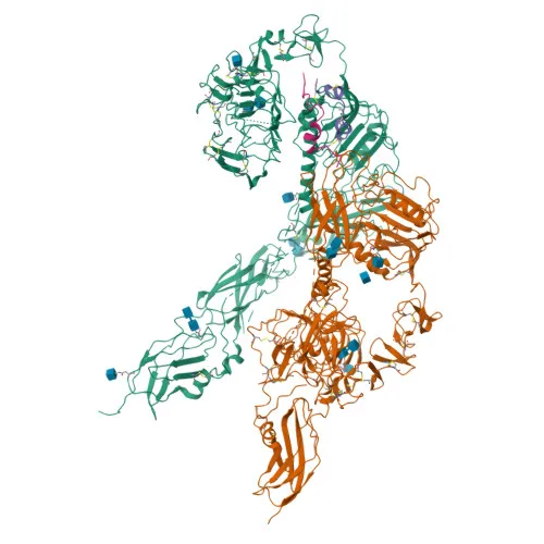
- IGF1R possesses key structural features including a tyrosine kinase domain, ATP-binding site, and disulfide bonds, and undergoes autophosphorylation on tyrosine residues upon activation.
Experimental Guidance and Technical Tips
- The immunogen is a synthetic phospho-peptide corresponding to residues surrounding tyrosine 1280 of human IGF1R, designed to detect phosphorylation at this site. Consider using phosphatase-treated controls to confirm phospho-specificity.
- This antibody is intended for Western blot (WB) detection. For optimal results, consider using whole-cell lysates from IGF1R-positive and stimulated cell lines, such as cancer lines known to overexpress IGF1R.
- Cross-reactivity is reported for human. If used with other species, validate reactivity experimentally before routine use.
- The predicted molecular weight is approximately 155 kDa. The phosphorylated form may exhibit a slight shift in migration; optimize gel conditions accordingly.
CamelBio: Your One-Stop Sourcing Bridge
As a trusted IVD raw-material partner, CamelBio provides high-quality antibodies like this phospho-IGF1R (Y1280) antibody, supported by technical expertise for your assay development. Whether you need validated antibody pairs, optimized polyclonal antibodies, or bulk ancillary reagents, we streamline procurement for cancer biomarker and signaling research. Reach out to discuss sourcing rare detection targets and accelerating your diagnostic workflows from concept to clinic.
Product Datasheet
Anti-Phospho-IGF1R (Y1280) Rabbit Polyclonal Antibody for WB - P08069
REQUEST A QUOTE
Our professional team will reply to you within one business day. Please feel free to contact us!