Phosphorylated Antibodies
Anti-Phospho-FLT3-Y589/591 Rabbit Polyclonal Antibody for WB, ELISA - P36888
Item Number : CM0001666
Price varies based on specs and customizations
- Application
- WB, ELISA
- Cross Reactivity
- Human
- Protein Weight
- 113kDa

Shipping:
Contact us to get shipping details Enjoy On-time Dispatch Guarantee.
Why Choose Us
Easy ordering process, quality products, and dedicated support for your business success.
Core Product Specifications and Parameters
| Parameter | Value |
|---|---|
| Product Name | Phospho-FLT3-Y589/591 Rabbit pAb |
| Remarks/Alias | FLK2; STK1; CD135; FLK-2; Phospho-FLT3-Y589/591 |
| Species | Human |
| Gene ID (Human) | 2322 |
| Gene ID | 2322 |
| Immunogen | Synthetic peptide | A synthetic phosphorylated peptide around Y589 & Y591 of human FLT3 (NP_004110.2). |
| Source | Rabbit |
| Category | Phosphorylated Antibodies |
| Application | WB, ELISA |
| Cross Reactivity | Human |
| SWISS | P36888 |
| Protein Weight | 113kDa |
| Shipping | Ice bag |
Biological Background: FLT3 Function and Localization
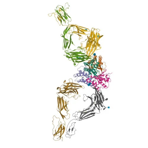
- FLT3 (Fms-like tyrosine kinase 3), also designated FLK2, STK1, and CD135, is a receptor-type tyrosine-protein kinase that binds FLT3LG to regulate differentiation, proliferation, and survival of hematopoietic progenitor cells and dendritic cells.
- Downstream signaling includes phosphorylation of SHC1 and AKT1, activation of MTOR, the RAS/MAPK cascade (MAPK1/ERK2, MAPK3/ERK1), and STAT5 proteins, though wild-type FLT3 induces only marginal STAT5 activation.
- FLT3 is located on the cell membrane and within the endoplasmic reticulum lumen.
- Expression is detected in bone marrow, hematopoietic stem cells, myeloid and granulocyte/macrophage progenitor cells; additionally in liver, thymus, spleen, lymph node, kidney, and pancreas, with high levels in T-cell leukemia.
- Mutations causing constitutive kinase activity drive cell proliferation and resistance to apoptosis via multiple pathways, highlighting FLT3 as a proto-oncogene and a key disease variant in acute myeloid leukemia.
- As a glycoprotein and phosphoprotein, FLT3 undergoes alternative splicing and is subject to ubiquitin conjugation; it also plays a role in host-virus interactions.
Experimental Guidance and Technical Tips
- Because this antibody targets phosphorylation sites, use lysis buffers supplemented with phosphatase inhibitors to preserve the phospho-epitope during sample preparation for Western blot.
- Consider stimulating FLT3-expressing cells (e.g., leukemia lines) with FLT3 ligand, or treating with kinase inhibitors, to confirm phosphorylation-specific detection.
- For ELISA, the immunogen phosphopeptide can serve as a positive control; plate coating and detection conditions may require optimization.
- Validate antibody specificity by comparing signals from wild-type and phosphatase-treated lysates or using site-directed mutagenesis of Y589/Y591.
CamelBio: Your One-Stop Sourcing Bridge
CamelBio is your one-stop partner for IVD raw-material sourcing, offering validated phosphorylation-specific antibodies, recombinant proteins, and ancillary reagents. For researchers studying FLT3-driven leukemia and kinase signaling, we provide reliable antibody pairs and bulk raw materials to accelerate diagnostic development. With comprehensive technical and supply chain support, CamelBio bridges the gap from target discovery to clinical application.
REQUEST A QUOTE
Our professional team will reply to you within one business day. Please feel free to contact us!