Phosphorylated Antibodies
Anti-Phospho-Chk2-T68 Rabbit Polyclonal Antibody for WB, ELISA - O96017
Item Number : CM0000772
Price varies based on specs and customizations
- Application
- WB, ELISA
- Cross Reactivity
- Human
- Protein Weight
- 61kDa

Shipping:
Contact us to get shipping details Enjoy On-time Dispatch Guarantee.
Why Choose Us
Easy ordering process, quality products, and dedicated support for your business success.
Core Product Specifications and Parameters
| Parameter | Details |
|---|---|
| Product Name | Phospho-Chk2-T68 Rabbit pAb |
| Remarks/Alias | CDS1; CHK2; LFS2; RAD53; hCds1; HuCds1; PP1425; Phospho-Chk2-T68 |
| Species | Human |
| Gene ID | 11200 |
| Immunogen | Synthetic phosphorylated peptide around T68 of human Chk2 |
| Source | Rabbit |
| Category | Phosphorylated Antibodies |
| Application | WB, ELISA |
| Cross Reactivity | Human |
| SWISS | O96017 |
| Protein Weight | 61kDa |
| Shipping | Ice bag |
Biological Background: Chk2 Function and Localization
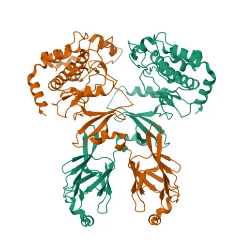
- Serine/threonine-protein kinase Chk2 (CHEK2, CDS1, RAD53 homolog) plays a critical role in checkpoint-mediated cell cycle arrest, DNA repair, and apoptosis in response to DNA double-strand breaks.
- Acts as a tumor suppressor; also negatively regulates cell cycle progression during unperturbed cycles and may contribute to chromosomal instability when absent.
- Following activation, phosphorylates numerous effectors preferentially at the consensus sequence [L-X-R-X-X-S/T].
Related references: PMID:37943659 - Regulates cell cycle checkpoint arrest by phosphorylating and inhibiting CDC25A, CDC25B, and CDC25C phosphatases, leading to inhibitory tyrosine phosphorylation of CDK-cyclin complexes.
- Promotes DNA repair through phosphorylation of BRCA2 (enhancing RAD51 chromatin association) and by stimulating DNA repair gene transcription via FOXM1.
- Mediates apoptosis through phosphorylation of p53/TP53 at Ser-20, MDM4, and PML, as well as through E2F1-dependent transcription of pro-apoptotic genes.
- Under oxidative stress, phosphorylates TRIM32 at Ser-55 to promote ATG7 ubiquitination and autophagy assembly.
Related references: PMID:37943659 - Promotes the CCAR2-SIRT1 association and is required for CCAR2-mediated SIRT1 inhibition.
Related references: PMID:25361978 - Predominantly localizes to the nucleus (nucleoplasm, PML bodies); highly expressed in testis, spleen, colon, and peripheral blood leukocytes, with lower expression in other tissues.
- Contains kinase, ATP-binding and nucleotide-binding domains; classified as a phosphoprotein with roles in mitosis, transcription regulation, and host-virus interaction.
Experimental Guidance and Technical Tips
- The immunogen is a synthetic phosphorylated peptide around T68, suggesting specificity for the phosphorylated form; use appropriate phosphatase inhibitors during sample preparation to preserve phospho-signals.
- For Western blot, consider stimulating cells with DNA-damaging agents (e.g., etoposide, irradiation) to induce Chk2 phosphorylation before lysate preparation.
- Validate the antibody in human samples; background band(s) may appear, and it is advisable to optimize loading and detection conditions.
- For ELISA, the phosphorylated peptide can serve as a coating antigen to confirm specificity; negative controls using non-phosphorylated peptide are recommended.
- An expected band near 61 kDa should be observed in WB; additional bands may reflect splice variants or post-translational modifications.
CamelBio: Your One-Stop Sourcing Bridge
CamelBio streamlines the sourcing of IVD raw materials from concept to clinic, offering diagnostic manufacturers and labs rapid access to this anti-phospho-Chk2 antibody for DNA damage and checkpoint kinase research. Beyond individual clones, our portfolio includes validated antibody pairs, optimized monoclonal/polyclonal antibodies, ancillary bulk reagents, and rare target sourcing—empowering your assay development with flexible, integrated supply solutions.
REQUEST A QUOTE
Our professional team will reply to you within one business day. Please feel free to contact us!