Phosphorylated Antibodies
Anti-Phospho-Akt-S473 Rabbit Monoclonal Antibody for WB, IHC-P, ELISA - P31749 / P31751 / Q9Y243
Item Number : CM0001927
Price varies based on specs and customizations
- Application
- WB, IHC-P, ELISA
- Cross Reactivity
- Human, Mouse, Rat

Shipping:
Contact us to get shipping details Enjoy On-time Dispatch Guarantee.
Why Choose Us
Easy ordering process, quality products, and dedicated support for your business success.
Core Product Specifications and Parameters
| Parameter | Value |
|---|---|
| Product Name | Phospho-Akt-S473 Rabbit mAb |
| Remarks/Alias | AKT1/AKT2/AKT3; Phospho-Akt-S473 |
| Species | Human |
| GeneID (human) | 207/208/10000 |
| GeneID | 207/208/10000 |
| Immunogen | Synthetic peptide; A synthetic phosphorylated peptide around S473 of human AKT1 (NP_005154.2) |
| Source | Rabbit |
| Category | Phosphorylated Antibodies |
| Application | WB, IHC-P, ELISA |
| Cross Reactivity | Human, Mouse, Rat |
| SWISS | P31749/P31751/Q9Y243 |
| Protein Weight | 48kDa/55kDa/51kDa/54kDa |
| Shipping | Ice bag |
Biological Background: AKT1 Function and Localization
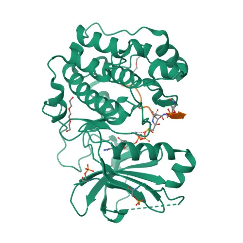
- AKT1 (RAC-alpha serine/threonine-protein kinase) is a member of the AKT kinase family and regulates metabolism, proliferation, cell survival, growth, and angiogenesis by phosphorylating over 100 downstream substrates. Related references: PMID:11882383 PMID:15526160 PMID:15861136 PMID:21432781
- It mediates insulin-induced glucose uptake via SLC2A4/GLUT4 translocation and stimulates glycogen synthesis by inhibiting GSK3 through phosphorylation.
- AKT1 promotes cell survival by phosphorylating and inhibiting pro-apoptotic proteins, including MAP3K5/ASK1 and BAD. Related references: PMID:11154276 PMID:10926925
- It activates the mTORC1 signaling pathway by phosphorylating TSC2, leading to increased protein synthesis and cell growth. Related references: PMID:12150915 PMID:12172553
- AKT1 localizes to the cytoplasm, nucleus, cell membrane, and mitochondrion intermembrane space, enabling diverse signaling roles.
- Expression is elevated in prostate and breast cancers, and the Tyr-176 phosphorylated form correlates with tumor progression from normal to metastatic stages.
- Post-translational modifications include phosphorylation at Thr-308 and Ser-473 for full activation, acetylation, ubiquitination, and disulfide bond formation, as noted in UniProt keywords.
- The antibody targets phosphorylated Ser-473, a critical activation site conserved across AKT1 (P31749), AKT2 (P31751), and AKT3 (Q9Y243).
Experimental Guidance and Technical Tips
- The immunogen is a synthetic phosphorylated peptide around Ser-473 of human AKT1; this design ensures high specificity for the phosphorylated epitope.
- For Western blot, include phosphatase inhibitors in the lysis buffer and consider using a low‑percentage BSA block to preserve phospho‑signals.
- For IHC‑P, optimize antigen retrieval (e.g., citrate buffer pH 6.0) and validate staining in samples with known AKT pathway activation, such as PTEN‑deficient tumors.
- For ELISA, the antibody can serve as a capture or detection reagent; perform cross‑titration experiments to confirm compatibility with your assay format and buffer conditions.
- Always include appropriate positive (e.g., insulin‑ or growth‑factor‑treated cell lysates) and negative (untreated or phosphatase‑treated) controls to verify phospho‑specificity.
CamelBio: Your One-Stop Sourcing Bridge
CamelBio simplifies the procurement of high‑quality IVD raw materials, offering validated phospho‑specific antibodies like this anti‑Phospho‑Akt‑S473 clone for oncology, metabolism, and signal transduction research. Our portfolio includes matched antibody pairs, optimized monoclonal and polyclonal reagents, bulk ancillary materials, and customized sourcing for rare‑target raw materials, enabling you to accelerate diagnostic assay development from concept to clinic.
Product Datasheet
Anti-Phospho-Akt-S473 Rabbit Monoclonal Antibody for WB, IHC-P, ELISA - P31749 / P31751 / Q9Y243
REQUEST A QUOTE
Our professional team will reply to you within one business day. Please feel free to contact us!