Monoclonal Antibodies
Anti-PHGDH Monoclonal Antibody for WB, IHC-P, IF/ICC, ELISA - O43175
Item Number : CM0004220
Price varies based on specs and customizations
- Application
- WB, IHC-P, IF/ICC, ELISA
- Cross Reactivity
- Human, Mouse, Rat
- Protein Weight
- 57kDa

Shipping:
Contact us to get shipping details Enjoy On-time Dispatch Guarantee.
Why Choose Us
Easy ordering process, quality products, and dedicated support for your business success.
Core Product Specifications and Parameters
| Parameter | Value |
|---|---|
| Product Name | PHGDH Rabbit mAb |
| Remarks/Alias | NLS; PDG; PGD; NLS1; PGAD; PGDH; SERA; 3PGDH; 3-PGDH; PHGDHD; HEL-S-113; PHGDH |
| Species | Human |
| Gene ID (Human) | 26227 |
| Immunogen | Recombinant fusion protein containing a sequence corresponding to amino acids 264-533 of human PHGDH (NP_006614.2) |
| Source | Rabbit |
| Category | Monoclonal Antibodies |
| Application | WB, IHC-P, IF/ICC, ELISA |
| Cross Reactivity | Human, Mouse, Rat |
| SWISS | O43175 |
| Protein Weight | 57kDa |
| Shipping | Ice bag |
Biological Background: PHGDH Function and Localization
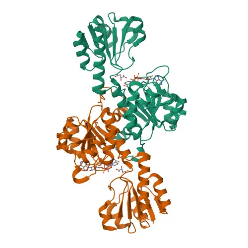
- PHGDH (D-3-phosphoglycerate dehydrogenase) is also known as 2-oxoglutarate reductase and malate dehydrogenase, reflecting its multi-substrate specificity.
- It catalyzes the reversible oxidation of 3-phospho-D-glycerate to 3-phosphonooxypyruvate, the first and rate-limiting step in the phosphorylated L-serine biosynthesis pathway.
- The enzyme also exhibits 2-oxoglutarate reductase and malate dehydrogenase activities, linking it to broader metabolic networks beyond serine synthesis.
- Post-translational modifications, including acetylation and phosphorylation, have been identified on PHGDH and may regulate its catalytic function or stability.
- UniProt keywords annotate PHGDH with roles in amino-acid biosynthesis, oxidoreductase activity, and reference proteome status, underscoring its conserved biological importance.
- No specific subcellular localization or tissue distribution data are provided in the curated record, but PHGDH is typically cytosolic in human cells.
Experimental Guidance and Technical Tips
- The immunogen covers amino acids 264–533 of human PHGDH; consider verifying the antibody’s recognition of full-length protein in your target sample system, especially for mouse and rat orthologs.
- For western blotting, expect a band near 57 kDa; optimize loading and detection conditions, and include positive and negative lysate controls.
- In IHC-P, test antigen retrieval methods (e.g., citrate vs. EDTA buffer) and confirm staining specificity using known PHGDH-expressing tissues.
- For IF/ICC, adjust fixation and permeabilization protocols for your cell type; co-staining with organelle markers can help confirm localization.
- ELISA applications may require coating the recombinant protein or cell lysate; include appropriate blanks and standard curves for quantitative analysis.
CamelBio: Your One-Stop Sourcing Bridge
CamelBio provides diagnostic manufacturers, laboratories, and research institutes with one-stop access to validated IVD raw materials, technical services, and consulting—covering every stage from concept to clinic. Our PHGDH monoclonal antibody supports research into serine metabolism and related pathologies. Beyond this product, CamelBio can supply optimized monoclonal/polyclonal antibodies, validated antibody pairs, bulk ancillary reagents, and assist with sourcing rare-target raw materials to accelerate your assay development.
REQUEST A QUOTE
Our professional team will reply to you within one business day. Please feel free to contact us!