Polyclonal Antibodies
Anti-PHD2/EGLN1 Polyclonal Antibody for WB, IHC-P, IF/ICC, ELISA - Q9GZT9
Item Number : CM0018045
Price varies based on specs and customizations
- Application
- WB, IHC-P, IF/ICC, ELISA
- Cross Reactivity
- Human, Mouse, Rat
- Protein Weight
- 46kDa

Shipping:
Contact us to get shipping details Enjoy On-time Dispatch Guarantee.
Why Choose Us
Easy ordering process, quality products, and dedicated support for your business success.
Core Product Specifications and Parameters
| Parameter | Value |
|---|---|
| Product Name | PHD2/EGLN1 Rabbit pAb |
| Remarks/Alias | HPH2; PHD2; SM20; ECYT3; HALAH; HPH-2; HIFPH2; ZMYND6; C1orf12; HIF-PH2; PHD2/EGLN1 |
| Species | Human |
| GeneID (Human) | 54583 |
| GeneID | 54583 |
| Immunogen | Recombinant fusion protein containing a sequence corresponding to amino acids 1-426 of human PHD2/EGLN1 (NP_071334.1) |
| Source | Rabbit |
| Category | Polyclonal Antibodies |
| Application | WB, IHC-P, IF/ICC, ELISA |
| Cross Reactivity | Human, Mouse, Rat |
| SWISS | Q9GZT9 |
| Protein Weight | 46kDa |
| Shipping | Ice bag |
Biological Background: PHD2/EGLN1 Function and Localization
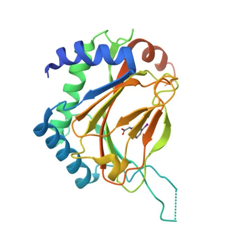
- PHD2/EGLN1 (Egl nine homolog 1), also designated HIF-prolyl hydroxylase 2 (HIF-PH2) or PHD2, is a cellular oxygen sensor encoded by the EGLN1 gene. It belongs to the EGLN family of prolyl hydroxylases and is the primary isozyme regulating hypoxia-inducible factor (HIF) stability under normoxic conditions.
- Under normal oxygen levels, PHD2 catalyzes the hydroxylation of specific proline residues within the oxygen-dependent degradation (ODD) domains of HIF-1α and HIF-2α. This post-translational modification promotes recognition by the von Hippel-Lindau (VHL) E3 ubiquitin ligase complex, leading to proteasomal degradation of HIFα subunits. Consequently, HIF-mediated transcriptional programs are suppressed.
- During hypoxia, PHD2 activity is diminished, allowing HIFα proteins to accumulate, dimerize with HIF-1β, and translocate to the nucleus to drive the expression of genes involved in angiogenesis, erythropoiesis, metabolic adaptation, and cell survival. This mechanism underlies cellular adaptation to reduced oxygen availability.
- PHD2 localizes predominantly to the cytoplasm but can also be detected in the nucleus, consistent with its role in sensing oxygen and modifying HIFα proteins in both compartments.
- Widely expressed across tissues, with highest mRNA levels observed in skeletal muscle, heart, brain, kidney, and adrenal gland (PubMed:11056053; PubMed:12351678). Protein is detected in cardiac myocytes, aortic endothelial cells, and coronary artery smooth muscle. Fetal tissues also show significant expression, particularly in brain, heart, spleen, and skeletal muscle (PubMed:12788921). Related references: PMID:11056053 PMID:12351678 PMID:12788921
- PHD2 undergoes several post-translational modifications including acetylation, phosphorylation, and S-nitrosylation, which may modulate its activity or stability. It requires iron, 2-oxoglutarate, and ascorbate (vitamin C) as cofactors for its dioxygenase activity, and contains a zinc-finger domain.
- Mutations in EGLN1 are associated with congenital erythrocytosis type 3 (ECYT3), highlighting its critical role in erythropoietin regulation and red blood cell production.
Experimental Guidance and Technical Tips
- The immunogen used to raise this polyclonal antibody is a recombinant protein encompassing almost the full length (aa 1-426) of human PHD2/EGLN1, corresponding to the canonical sequence. This design may favor recognition of the endogenous protein across multiple applications.
- For Western blotting (WB) applications, a specific band corresponding to the predicted molecular weight of approximately 46 kDa should be observed. Optimization of sample preparation, loading amount, and detection conditions is recommended. Positive controls derived from tissues with high PHD2 expression, such as skeletal muscle or heart, may be beneficial.
- In immunohistochemistry (IHC-P) and immunofluorescence (IF/ICC), consider testing a range of antigen retrieval methods and antibody dilutions. Given the dual cytoplasmic/nuclear localization of PHD2, staining patterns should be evaluated in the context of the cellular oxygen status. Appropriate tissue type or cell line controls (e.g., normoxic vs. hypoxic) can aid in confirming specificity.
- For ELISA-based assays, the antibody may be suitable as a capture or detection reagent after validation in the relevant sample matrix. Users should perform cross-reactivity and interference testing according to their specific protocol requirements.
- While this antibody is reported to cross-react with mouse and rat PHD2/EGLN1, sequence alignment and pilot experiments are advised to confirm equivalent reactivity in these species for the intended application.
CamelBio: Your One-Stop Sourcing Bridge
CamelBio serves as a comprehensive sourcing partner for IVD manufacturers and research laboratories, offering a broad portfolio of qualified raw materials, from antibodies and recombinant proteins to ancillary reagents. This anti-PHD2/EGLN1 polyclonal antibody supports investigations into hypoxia signaling, angiogenesis, and oncology biomarker discovery, and exemplifies the quality of tools available through our platform. Whether you require validated antibody pairs, bulk monoclonal or polyclonal antibodies, or assistance sourcing hard-to-find targets, CamelBio streamlines your procurement process to accelerate assay development from concept to clinical application.
Product Datasheet
Anti-PHD2/EGLN1 Polyclonal Antibody for WB, IHC-P, IF/ICC, ELISA - Q9GZT9
REQUEST A QUOTE
Our professional team will reply to you within one business day. Please feel free to contact us!