Polyclonal Antibodies
Anti-PGE receptor EP3 (PTGER3) Rabbit Polyclonal Antibody for WB, ELISA - P43115
Item Number : CM0027744
Price varies based on specs and customizations
- Application
- WB, ELISA
- Cross Reactivity
- Mouse, Rat
- Protein Weight
- 43kDa

Shipping:
Contact us to get shipping details Enjoy On-time Dispatch Guarantee.
Why Choose Us
Easy ordering process, quality products, and dedicated support for your business success.
Core Product Specifications and Parameters
| Parameter | Value |
|---|---|
| Product Name | PGE receptor EP3 (PTGER3) Rabbit pAb |
| Remarks/Alias | EP3; EP3e; EP3-I; EP3-II; EP3-IV; EP3-VI; PGE2-R; EP3-III; lnc003875; PGE receptor EP3 (PTGER3) |
| Species | Human |
| GeneID (Human) | 5733 |
| GeneID | 5733 |
| Immunogen | Recombinant fusion protein containing a sequence corresponding to amino acids 1-53 of human PGE receptor EP3 (PTGER3) (NP_942009.1) |
| Source | Rabbit |
| Category | Polyclonal Antibodies |
| Application | WB, ELISA |
| Cross Reactivity | Mouse, Rat |
| SWISS | P43115 |
| Protein Weight | 43kDa |
| Shipping | Ice bag |
Biological Background: PTGER3 Function and Localization
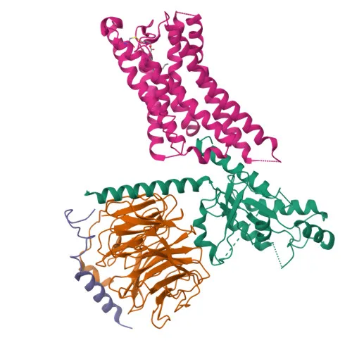
- PTGER3 encodes the Prostaglandin E2 receptor EP3 subtype (PGE2-R, prostanoid EP3 receptor), a G protein-coupled receptor (GPCR) belonging to the rhodopsin-like receptor family. Related references: PMID:7883006 PMID:7981210 PMID:8117308
- Alternative splicing generates multiple isoforms (EP3, EP3e, EP3-I, EP3-II, EP3-IV, EP3-VI, EP3-III) that may differ in C-terminal tails and signaling properties.
- The receptor couples to G(i) proteins to inhibit adenylate cyclase and can also elevate intracellular calcium, mediating diverse physiological responses to PGE2.
- PTGER3 plays essential roles in fever development (response to pyrogens like IL1B and LPS), platelet aggregation potentiation, and duodenal mucosal protection via increased bicarbonate secretion.
- Subcellular localization is primarily at the cell membrane, consistent with its function as a transmembrane receptor.
- Tissue expression includes kidney, small intestine, heart, pancreas, gastric fundic mucosa, mammary artery, and pulmonary vessels. Related references: PMID:8117308 PMID:8135729
- As a membrane protein, PTGER3 is subject to post-translational modifications including glycosylation and palmitoylation, which may regulate receptor stability and signaling.
Experimental Guidance and Technical Tips
- The immunogen corresponds to the N-terminal region (aa 1-53) of human PTGER3, which is conserved across human, mouse, and rat, supporting the observed cross-reactivity.
- For Western blotting, the predicted molecular weight is 43kDa; optimize gel percentage and transfer conditions accordingly.
- Validate antibody specificity in positive control tissues such as kidney or small intestine, where PTGER3 expression is well-documented.
- This polyclonal antibody may detect multiple isoforms due to alternative splicing; confirm band patterns using appropriate controls.
- In ELISA applications, consider using recombinant protein as standard to establish assay sensitivity and specificity.
CamelBio: Your One-Stop Sourcing Bridge
At CamelBio, we provide diagnostic manufacturers and research labs with a one-stop procurement solution for IVD raw materials, from concept to clinic. This anti-PTGER3 antibody supports studies in prostaglandin signaling, inflammation, and platelet function, and we can further source validated antibody pairs, bulk ancillary reagents, and rare-target raw materials to accelerate your assay development. Whether you need optimized polyclonal antibodies or customized sourcing, CamelBio bridges the gap between discovery and scalable supply.
Product Datasheet
Anti-PGE receptor EP3 (PTGER3) Rabbit Polyclonal Antibody for WB, ELISA - P43115
REQUEST A QUOTE
Our professional team will reply to you within one business day. Please feel free to contact us!