Monoclonal Antibodies
Anti-PERK Rabbit Monoclonal Antibody for WB, ELISA - Q9NZJ5
Item Number : CM0008831
Price varies based on specs and customizations
- Application
- WB, ELISA
- Cross Reactivity
- Human, Mouse, Rat
- Protein Weight
- 125kDa

Shipping:
Contact us to get shipping details Enjoy On-time Dispatch Guarantee.
Why Choose Us
Easy ordering process, quality products, and dedicated support for your business success.
Core Product Specifications and Parameters
| Parameter | Value |
|---|---|
| Product Name | PERK Rabbit mAb |
| Remarks/Alias | PEK; WRS; PERK |
| Species | Human |
| GeneID (Human) | 9451 |
| GeneID | 9451 |
| Immunogen | Recombinant protein |
| Source | Rabbit |
| Category | Monoclonal Antibodies |
| Application | WB, ELISA |
| Cross Reactivity | Human, Mouse, Rat |
| SWISS | Q9NZJ5 |
| Protein Weight | 125kDa |
| Shipping | Ice bag |
Biological Background: PERK Function and Localization
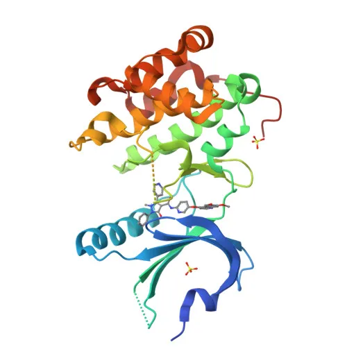
- Eukaryotic translation initiation factor 2-alpha kinase 3 (PERK), also known as PEK and HsPEK, is a stress-sensing protein kinase that plays a central role in the unfolded protein response (UPR) by phosphorylating the alpha subunit of translation initiation factor eIF2 (eIF2α). Related references: PMID:10026192, PMID:10677345, PMID:11907036
- Upon recognizing misfolded proteins in the endoplasmic reticulum, PERK activation leads to eIF2α phosphorylation, resulting in global attenuation of cap-dependent translation while selectively enhancing translation of stress-responsive mRNAs such as ATF4 and QRICH1. Related references: PMID:10026192, PMID:10677345, PMID:27917829, PMID:31023583
- PERK also phosphorylates NRF2, promoting its nuclear accumulation and activation of antioxidant response pathways.
- PERK-mediated UPR increases mitochondrial oxidative phosphorylation and respiratory chain supercomplex formation, while mitochondrial function is protected at ER–mitochondria contact sites by ATAD3A-mediated PERK inhibition. Related references: PMID:31023583, PMID:39116259
- PERK signaling contributes to UPR-induced G1 cell cycle arrest through the loss of cyclin D1.
- PERK is localized to the endoplasmic reticulum membrane.
- Ubiquitously expressed with high levels in secretory tissues.
Experimental Guidance and Technical Tips
- The immunogen used is recombinant protein; consider using a recombinant PERK protein as a positive control in your assay to verify antibody specificity.
- For Western blot, the expected molecular weight is approximately 125 kDa. Ensure appropriate protein extraction and loading conditions, and validate results using relevant cellular models of ER stress.
- For ELISA applications, titrate the antibody to determine the optimal working concentration in your specific assay format, and include appropriate negative and positive controls.
CamelBio: Your One-Stop Sourcing Bridge
CamelBio offers a comprehensive one-stop platform for IVD raw materials, supporting diagnostic manufacturers and research laboratories with validated antibody pairs, monoclonal/polyclonal antibodies, and bulk ancillary reagents. This PERK antibody supports research in stress biology, unfolded protein response, and related disease areas. For custom sourcing needs, technical consultation, or large-scale procurement, our team is ready to assist.
REQUEST A QUOTE
Our professional team will reply to you within one business day. Please feel free to contact us!