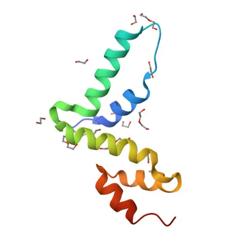
Polyclonal Antibodies
Anti-PEG3 Polyclonal Antibody for WB, IHC-P, ELISA - Q9GZU2
Item Number : CM0016955
Price varies based on specs and customizations
- Application
- WB, IHC-P, ELISA
- Cross Reactivity
- Human, Mouse, Rat
- Protein Weight
- 181kDa

Shipping:
Contact us to get shipping details Enjoy On-time Dispatch Guarantee.
Why Choose Us
Easy ordering process, quality products, and dedicated support for your business success.
Core Product Specifications and Parameters
| Parameter | Value |
|---|---|
| Product Name | PEG3 Rabbit pAb |
| Remarks/Alias | PW1; ZNF904; ZSCAN24; ZKSCAN22; PEG3 |
| Species | Human |
| Gene ID (Human) | 5178 |
| Gene ID | 5178 |
| Immunogen | Recombinant protein |
| Source | Rabbit |
| Category | Polyclonal Antibodies |
| Application | WB, IHC-P, ELISA |
| Cross Reactivity | Human, Mouse, Rat |
| SWISS | Q9GZU2 |
| Protein Weight | 181kDa |
| Shipping | Ice bag |
Biological Background: PEG3 Function and Localization
- Paternally-expressed gene 3 protein (PEG3), also known as Zinc finger and SCAN domain-containing protein 24 (ZSCAN24) and ZNF904, belongs to the Krüppel C2H2-type zinc-finger protein family with SCAN box domain.
- PEG3 induces apoptosis in cooperation with SIAH1A and acts as a mediator between p53/TP53 and BAX in the neuronal death pathway activated by DNA damage.
- It acts synergistically with TRAF2 and inhibits TNF-induced apoptosis through activation of NF-kappa-B, demonstrating a dual role in cell death regulation.
- PEG3 possesses tumor suppressing activity in glioma cells, with expression highly repressed in glioma cell lines, suggesting a role in cancer pathogenesis.
- The protein localizes to both the nucleus and cytoplasm, consistent with its transcriptional regulatory and signaling functions.
- Tissue expression is enriched in brain, glial cells, astrocytes, embryo, placenta, testis, ovary, and uterus. In placenta, it is found in villous cytotrophoblast cells, while in ovary it is present in stromal cells including thecal layers around follicles.
- Key post-translational modifications and structural features include metal-binding, alternative splicing, and multiple zinc-finger repeats, all of which are critical for its diverse biological activities.
Experimental Guidance and Technical Tips
- The immunogen corresponds to amino acids 195-400, a central region of PEG3; this design may afford recognition of core functional domains and could enable detection across splice variants or orthologs.
- For Western blotting, consider using whole cell lysates from brain or placental tissues where PEG3 is highly expressed; a band near 181kDa is expected, so optimize gel percentage and transfer conditions for high molecular weight proteins.
- For immunohistochemistry (IHC-P), test on formalin-fixed paraffin-embedded human brain or placental sections; antigen retrieval may be required, and appropriate controls should be included.
- The antibody cross-reacts with mouse and rat PEG3; validate in the relevant model system using positive tissue controls (e.g., mouse brain, rat testis) to confirm specificity.
- For ELISA, the polyclonal nature may provide robust capture/detection; consider pairing with a monoclonal antibody if sensitivity is insufficient.
CamelBio: Your One-Stop Sourcing Bridge
CamelBio supports diagnostic manufacturers and researchers by providing reliable raw materials for apoptosis and tumor biology projects. This PEG3 polyclonal antibody, suitable for WB, IHC-P, and ELISA, complements our portfolio of optimized monoclonal/polyclonal antibodies and ancillary reagents. From target discovery to assay development, our streamlined sourcing covers validated pairs, bulk supplies, and rare-target procurement—helping you bridge the gap from concept to clinic with confidence.
REQUEST A QUOTE
Our professional team will reply to you within one business day. Please feel free to contact us!