Polyclonal Antibodies
Anti-PDK1/PDPK1 Rabbit Polyclonal Antibody for WB and ELISA - O15530
Item Number : CM0023024
Price varies based on specs and customizations
- Application
- WB, ELISA
- Cross Reactivity
- Human, Mouse, Rat
- Protein Weight
- 63kDa

Shipping:
Contact us to get shipping details Enjoy On-time Dispatch Guarantee.
Why Choose Us
Easy ordering process, quality products, and dedicated support for your business success.
Core Product Specifications and Parameters
| Parameter | Value |
|---|---|
| Product Name | PDK1/PDPK1 Rabbit pAb |
| Remarks/Alias | PDK1, PDPK2, PDPK2P, PRO0461, PDK1/PDPK1 |
| Species | Human |
| Gene ID (Human) | 5170 |
| Immunogen | Recombinant fusion protein containing a sequence corresponding to amino acids 409-556 of human PDK1/PDPK1 (NP_002604.1) |
| Source | Rabbit |
| Category | Polyclonal Antibodies |
| Application | WB, ELISA |
| Cross Reactivity | Human, Mouse, Rat |
| SWISS | O15530 |
| Protein Weight | 63kDa |
| Shipping | Ice bag |
Biological Background: PDK1/PDPK1 Function and Localization
-
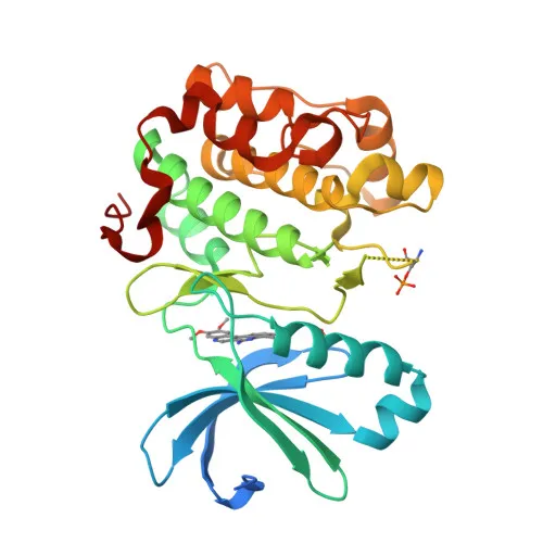
PDK1 (3-phosphoinositide-dependent protein kinase 1) is a serine/threonine kinase that functions as a master kinase for the AGC kinase family, phosphorylating and activating multiple downstream targets. Related references: PMID:10226025, PMID:10480933, PMID:10995762
-
It activates key signaling proteins such as AKT/PKB, p70 S6 kinase, p90 RSK, PKC isoforms, SGK, and PAK1, thereby playing a central role in insulin signal transduction, regulation of cell proliferation, survival, and metabolism. Related references: PMID:10226025, PMID:10480933, PMID:10995762, PMID:12167717, PMID:9094314
-
PDK1 negatively regulates TGF-beta signaling by modulating SMAD protein interactions and phosphorylation, preventing nuclear accumulation of SMAD3 and SMAD4. Related references: PMID:17327236
-
It activates the NF-kappa-B pathway via IKKB phosphorylation and regulates focal adhesion dynamics through tyrosine phosphorylation. Related references: PMID:16207722, PMID:14585963
-
PDK1 is essential for vascular endothelial cell motility and chemotaxis, participates in calcium entry and mast cell function, and plays critical roles in cardiac homeostasis and thymocyte development. Related references: PMID:17371830
-
Subcellularly, PDK1 localizes to the cytoplasm, nucleus, cell membrane, and focal adhesions, enabling its diverse signaling functions.
-
Post-translational modifications include acetylation, phosphorylation (notably Tyr-9, elevated in diseased tissues), and ubiquitin conjugation.
-
PDK1 is ubiquitously expressed, and Tyr-9 phosphorylated form is markedly increased in diseased lung, liver, colon, and breast tissues compared to normal.
Experimental Guidance and Technical Tips
- The immunogen corresponds to amino acids 409-556 of human PDK1, a region within the C-terminal portion of the kinase domain. Consider that the antibody may recognize both unphosphorylated and phosphorylated PDK1 forms; specific phosphorylation states should be validated independently.
- For Western blot, a band around 63 kDa is expected. Use appropriate loading controls and validate detection in human, mouse, or rat cell lysates. For ELISA, ensure optimal coating conditions and antibody titration.
- Given PDK1's role in insulin and growth factor signaling, consider stimulating cells with insulin or serum to assess pathway activation-related changes in PDK1 expression or phosphorylation.
CamelBio: Your One-Stop Sourcing Bridge
CamelBio provides one-stop access to high-quality IVD raw materials, including this anti-PDK1 rabbit polyclonal antibody, supporting research into insulin signaling, cancer biology, and related pathways. Our offerings encompass validated antibody pairs, optimized polyclonal and monoclonal antibodies, bulk ancillary reagents, and rare target raw-material sourcing, streamlining your diagnostic development from concept to clinic.
REQUEST A QUOTE
Our professional team will reply to you within one business day. Please feel free to contact us!