Monoclonal Antibodies
Anti-PDIA6 Rabbit Monoclonal Antibody for WB, IF/ICC, ELISA - Q15084
Item Number : CM0009676
Price varies based on specs and customizations
- Application
- WB, IF/ICC, ELISA
- Cross Reactivity
- Human, Mouse, Rat
- Protein Weight
- 48kDa

Shipping:
Contact us to get shipping details Enjoy On-time Dispatch Guarantee.
Why Choose Us
Easy ordering process, quality products, and dedicated support for your business success.
Core Product Specifications and Parameters
| Parameter | Value |
|---|---|
| Product Name | PDIA6 Rabbit mAb |
| Remarks/Alias | P5, ERP5, TXNDC7, PDIA6 |
| Species | Human |
| Gene ID | 10130 |
| Immunogen | A synthetic peptide corresponding to a sequence within amino acids 1-100 of human PDIA6 (NP_005733.1). |
| Source | Rabbit |
| Category | Monoclonal Antibodies |
| Application | WB, IF/ICC, ELISA |
| Cross Reactivity | Human, Mouse, Rat |
| SWISS | Q15084 |
| Protein Weight | 48kDa |
| Shipping | Ice bag |
Biological Background: PDIA6 Function and Localization
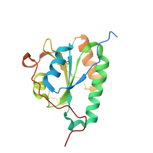
- PDIA6 (Protein disulfide-isomerase A6), also designated ERP5, P5, or TXNDC7, belongs to the protein disulfide isomerase family and contains thioredoxin domains.
- Functions as a molecular chaperone that inhibits the aggregation of misfolded proteins, contributing to proteostasis in the endoplasmic reticulum. Related references: PMID:12204115
- Negatively regulates the unfolded protein response (UPR) by binding to UPR sensors such as ERN1 (IRE1α) and EIF2AK3 (PERK), thereby attenuating their downstream signaling cascades. Related references: PMID:24508390
- Plays a role in platelet aggregation and activation upon stimulation by agonists including convulxin, collagen, and thrombin. Related references: PMID:15466936
- Localizes predominantly to the endoplasmic reticulum lumen, but is also detected at the cell membrane and in melanosomes, suggesting diverse functional niches.
- Expressed in platelets at the protein level; post-translational modifications include phosphorylation and disulfide bond formation, indicative of regulatory redox activity.
Experimental Guidance and Technical Tips
- The immunogen sequence (amino acids 1-100 of human PDIA6) corresponds to the N-terminal region; it may be suitable for detecting full-length protein in denaturing Western blots. For native applications like IF/ICC, epitope accessibility should be verified.
- In Western blot, expect a band near 48 kDa; validate detection in cell lines such as HEK293 or platelet lysates.
- For immunofluorescence, PDIA6 exhibits a reticular ER staining pattern; co-localization with an ER marker is recommended to confirm specificity.
- Stated cross-reactivity with mouse and rat should be verified in appropriate tissue, as sequence variations in the immunogen region could affect binding.
- For ELISA, consider using this antibody as a capture or detection reagent after titration and validation with recombinant PDIA6.
- To study PDIA6's role in UPR, ER stress induction (e.g., with tunicamycin) may modulate its expression.
- Use reducing conditions for electrophoresis to account for intramolecular disulfide bonds and achieve expected molecular weight.
CamelBio: Your One-Stop Sourcing Bridge
CamelBio provides diagnostic manufacturers, labs, and research institutes with one-stop access to IVD raw materials, technical services, and consulting—covering every stage from concept to clinic. As a trusted sourcing partner, CamelBio supports your PDIA6 research with validated antibody pairs, optimized monoclonal and polyclonal antibodies, bulk ancillary reagents, and rare target raw-material sourcing. Whether you aim to develop ER stress biomarkers or platelet activation assays, our comprehensive portfolio streamlines your assay development.
REQUEST A QUOTE
Our professional team will reply to you within one business day. Please feel free to contact us!