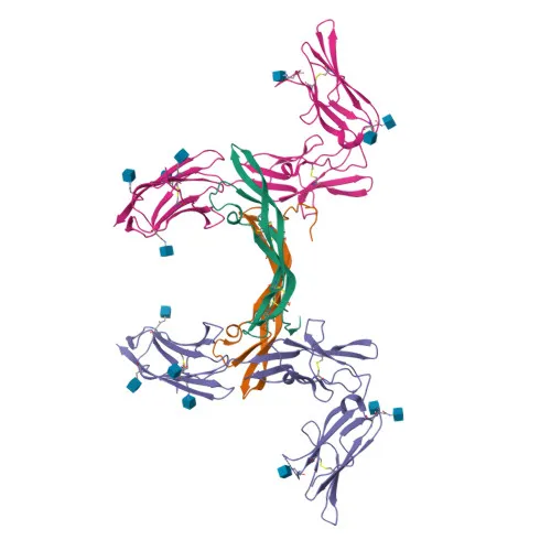
Polyclonal Antibodies
Anti-PDGFRB Polyclonal Antibody for WB - P09619
Item Number : CM0017703
Price varies based on specs and customizations
- Application
- WB, IHC-P, IF/ICC, ELISA
- Cross Reactivity
- Human, Mouse, Rat
- Protein Weight
- 124 kDa

Shipping:
Contact us to get shipping details Enjoy On-time Dispatch Guarantee.
Why Choose Us
Easy ordering process, quality products, and dedicated support for your business success.
Core Product Specifications and Parameters
| Parameter | Value |
|---|---|
| Product Name | PDGFRB Rabbit pAb |
| Remarks/Alias | IMF1; KOGS; IBGC4; JTK12; PDGFR; PENTT; CD140B; PDGFR1; PDGFR-1; PDGFRB |
| Species | Human |
| GeneID (Human) | 5159 |
| Gene ID | 5159 |
| Immunogen | Recombinant fusion protein containing a sequence corresponding to amino acids 900-1000 of human PDGFRB (NP_002600.1). |
| Source | Rabbit |
| Category | Polyclonal Antibodies |
| Application | WB, IHC-P, IF/ICC, ELISA |
| Cross Reactivity | Human, Mouse, Rat |
| SWISS | P09619 |
| Protein Weight | 124 kDa |
| Shipping | Ice bag |
Biological Background: PDGFRB Function and Localization
- PDGFRB encodes the platelet-derived growth factor receptor beta, a receptor tyrosine kinase that binds homodimeric PDGFB and PDGFD and heterodimers formed by PDGFA and PDGFB.
- It plays essential roles in embryonic development, cell proliferation, survival, differentiation, chemotaxis, and migration.
- PDGFRB is critical for blood vessel development, promoting proliferation, migration, and recruitment of pericytes and smooth muscle cells to endothelial cells; it also drives neointima formation at vascular injury sites.
- Required for normal kidney glomerulus development, it facilitates the formation of a branched capillary network and recruitment of mesangial cells.
- Ligand binding activates multiple signaling cascades, including PLCG1-mediated calcium mobilization, PI3K-AKT, and RAS-MAPK pathways, leading to cytoskeletal reorganization and membrane ruffling.
- The receptor is located at the cell membrane, cytoplasmic vesicles, and lysosome lumen; signaling is downregulated by dephosphorylation and rapid internalization.
- Post-translational modifications include disulfide bond formation, glycosylation, phosphorylation, and ubiquitination (Keywords: Glycoprotein, Phosphoprotein, Ubl conjugation). Alternative splicing has been reported.
Experimental Guidance and Technical Tips
- The immunogen corresponds to the C-terminal region (amino acids 900–1000) of human PDGFRB, which may detect full-length and potential cleaved or truncated forms.
- For Western blot (WB), the predicted molecular weight is 124 kDa; consider using sufficient protein loading and optimized transfer conditions due to the high molecular weight.
- In immunohistochemistry (IHC-P) and immunofluorescence (IF/ICC), validate the antibody in tissues or cells with known PDGFRB expression, such as vascular smooth muscle or kidney glomeruli.
- For ELISA, optimize coating antigen concentrations and include appropriate controls to ensure specificity.
- Cross-reactivity with mouse and rat suggests suitability for rodent model studies; however, verify sequence homology of the immunogen region in these species.
CamelBio: Your One-Stop Sourcing Bridge
CamelBio provides diagnostic manufacturers, labs, and research institutes with one-stop access to IVD raw materials, technical services, and consulting—covering every stage from concept to clinic. This anti-PDGFRB polyclonal antibody is ideal for oncology, cardiovascular, and renal disease research, serving as a robust component in assay development. Our portfolio includes validated antibody pairs, optimized monoclonal/polyclonal antibodies, bulk ancillary reagents, and rare target raw-material sourcing to streamline your IVD projects.
REQUEST A QUOTE
Our professional team will reply to you within one business day. Please feel free to contact us!