Polyclonal Antibodies
Anti-PDE4A Polyclonal Antibody for WB, IHC-P, IF/ICC, ELISA - P27815
Item Number : CM0019432
Price varies based on specs and customizations
- Application
- WB, IHC-P, IF/ICC, ELISA
- Cross Reactivity
- Human, Mouse, Rat
- Protein Weight
- 98kDa

Shipping:
Contact us to get shipping details Enjoy On-time Dispatch Guarantee.
Why Choose Us
Easy ordering process, quality products, and dedicated support for your business success.
Core Product Specifications and Parameters
| Parameter | Value |
|---|---|
| Product Name | PDE4A Rabbit pAb |
| Remarks/Alias | PDE4; DPDE2; PDE46; PDE4A |
| Species | Human |
| Gene ID (Human) | 5141 |
| Gene ID | 5141 |
| Immunogen | Recombinant fusion protein containing a sequence corresponding to amino acids 325-410 of human PDE4A (NP_001104777.1) |
| Source | Rabbit |
| Category | Polyclonal Antibodies |
| Application | WB, IHC-P, IF/ICC, ELISA |
| Cross Reactivity | Human, Mouse, Rat |
| SWISS | P27815 |
| Protein Weight | 98kDa |
| Shipping | Ice bag |
Biological Background: PDE4A Function and Localization
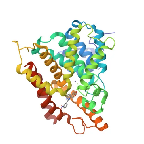
- PDE4A, also known as cAMP-specific phosphodiesterase 4A, is a member of the phosphodiesterase 4 family that specifically hydrolyzes the second messenger 3',5'-cyclic AMP (cAMP) to AMP, thereby downregulating cAMP signaling pathways.
- The enzyme plays a critical role in cell signaling by controlling intracellular cAMP concentrations, influencing processes such as inflammation, smooth muscle contraction, and cognitive function.
- PDE4A is characterized by its independence from calcium, calmodulin, and cGMP levels; it does not hydrolyze cGMP, distinguishing it from other phosphodiesterases.
- The gene undergoes alternative splicing, producing multiple isoforms with distinct regulatory features and tissue distributions.
- Subcellularly, PDE4A localizes to the cytoplasm, perinuclear region, cytosol, membrane, and cell projections such as ruffle membranes, indicating involvement in compartmentalized cAMP signaling.
- It is highly expressed in organs including liver, stomach, testis, thyroid, adrenal glands, heart, and small intestine, as well as in various immune cell subsets like T cells, dendritic cells, macrophages, and mast cells.
- Post-translational modifications include phosphorylation, ubiquitination, and isopeptide bond formation, with the protein binding manganese and zinc ions as cofactors.
Experimental Guidance and Technical Tips
- The immunogen sequence (amino acids 325-410) corresponds to a region within the catalytic domain; verify antibody specificity against desired isoforms using WB with recombinant proteins or cell lysates from relevant species.
- For WB, a band around 98 kDa is expected; consider using positive control lysates from tissues with high PDE4A expression (e.g., liver, heart) or from stimulated immune cells.
- In IHC-P and IF/ICC, include both negative and positive tissue controls (e.g., human or rodent tissues with known expression patterns) and optimize antigen retrieval conditions.
- ELISA applications may require careful titration to achieve optimal signal-to-noise ratio; cross-reactivity with mouse and rat supports multi-species assays but always validate in your specific experimental system.
CamelBio: Your One-Stop Sourcing Bridge
CamelBio offers diagnostic manufacturers and laboratories a streamlined gateway to IVD raw materials, providing not only high-quality antibodies like this PDE4A polyclonal but also validated antibody pairs, optimized monoclonal options, bulk ancillary reagents, and sourcing of rare targets. With expertise in cAMP signaling and phosphodiesterase research, CamelBio supports your projects from early-stage validation through to clinical application, backed by comprehensive technical and supply chain services.
REQUEST A QUOTE
Our professional team will reply to you within one business day. Please feel free to contact us!