Monoclonal Antibodies
Anti-PCK1 Monoclonal Antibody (PEPCK-C) for Western Blot and ELISA - P35558
Item Number : CM0006904
Price varies based on specs and customizations
- Application
- WB, ELISA
- Cross Reactivity
- Human, Mouse, Rat
- Protein Weight
- 69kDa

Shipping:
Contact us to get shipping details Enjoy On-time Dispatch Guarantee.
Why Choose Us
Easy ordering process, quality products, and dedicated support for your business success.
Core Product Specifications and Parameters
| Parameter | Value |
|---|---|
| Product Name | PCK1 Rabbit mAb |
| Remarks/Alias | PCKDC; PEPCK1; PEPCKC; PEPCK-C; PCK1 |
| Species | Human |
| Gene ID | 5105 |
| Immunogen | Synthetic peptide corresponding to a sequence within amino acids 523-622 of human PCK1 (NP_002582.3) |
| Source | Rabbit |
| Category | Monoclonal Antibodies |
| Application | WB, ELISA |
| Cross Reactivity | Human, Mouse, Rat |
| SWISS | P35558 |
| Protein Weight | 69kDa |
| Shipping | Ice bag |
Biological Background: PCK1 Function and Localization
-
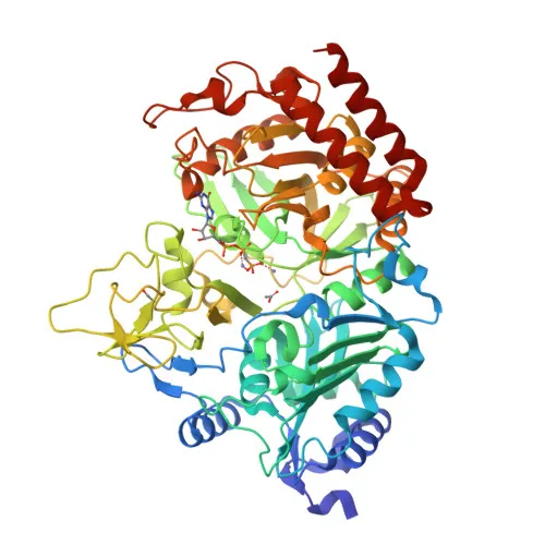
PCK1 (phosphoenolpyruvate carboxykinase 1) is the cytosolic isoform of PEPCK, serving as the rate-limiting enzyme of gluconeogenesis. It catalyzes the reversible decarboxylation and phosphorylation of oxaloacetate to phosphoenolpyruvate, balancing cataplerotic and anaplerotic fluxes in the citric acid cycle. Related references: PMID:24863970, PMID:26971250, PMID:28216384
-
Under low glucose conditions, PCK1 drives the cataplerotic conversion of oxaloacetate to phosphoenolpyruvate for gluconeogenesis; under high glucose, it promotes anaplerotic conversion of phosphoenolpyruvate to oxaloacetate. Related references: PMID:30193097
-
Beyond its metabolic function, phosphorylation at Ser-90 by AKT1 switches PCK1 to a serine-protein kinase. This moonlighting kinase activity triggers translocation to the endoplasmic reticulum, where it phosphorylates INSIG1/2 to release SREBP transcription factors and activate lipogenic gene expression. Related references: PMID:32322062
-
PCK1 contributes to memory CD8+ T-cell homeostasis by supplying phosphoenolpyruvate to glycogen and pentose phosphate pathways (by similarity).
-
Subcellularly, PCK1 is predominantly cytosolic, but upon AKT-mediated phosphorylation, it translocates to the endoplasmic reticulum.
-
Major tissue expression sites include the liver, kidney, and adipocytes, reflecting its central role in glucose and lipid metabolism.
-
Post-translational modifications include acetylation, ubiquitination, and phosphorylation, and structurally PCK1 is classified as a GTP-binding kinase, lyase, and decarboxylase.
Experimental Guidance and Technical Tips
- The immunogen was raised against a synthetic peptide spanning amino acids 523–622 near the C-terminus, distal to the Ser-90 phosphorylation site; consider this when designing phospho-specific or isoform-specific detection experiments.
- For Western blotting, validate detection in human, mouse, or rat liver/kidney lysates, as these tissues express high levels of PCK1. Induction of gluconeogenesis (e.g., fasting/cAMP treatment) may enhance signal.
- In ELISA, test antibody pairing and compatibility with sandwich formats; the monoclonal nature ensures consistent specificity across lots.
- Given the 69 kDa predicted molecular weight, ensure adequate gel resolution and transfer conditions; post-translational modifications may shift the observed band.
CamelBio: Your One-Stop Sourcing Bridge
CamelBio provides diagnostic manufacturers and metabolic research labs with one-stop access to validated IVD raw materials, including monoclonal and polyclonal antibodies like this PCK1 clone. From bulk ancillary reagents to rare target raw-material sourcing, we support your entire assay development workflow—helping you move seamlessly from concept to clinic. For gluconeogenesis, diabetes, or lipid metabolism studies, our PCK1 antibody supply can be combined with optimized antibody pairs and technical consultation.
REQUEST A QUOTE
Our professional team will reply to you within one business day. Please feel free to contact us!