Polyclonal Antibodies
Anti-PAK4 Rabbit Polyclonal Antibody for WB, IF/ICC, ELISA - O96013
Item Number : CM0015673
Price varies based on specs and customizations
- Application
- WB, IF/ICC, ELISA
- Cross Reactivity
- Human, Mouse
- Protein Weight
- 64kDa

Shipping:
Contact us to get shipping details Enjoy On-time Dispatch Guarantee.
Why Choose Us
Easy ordering process, quality products, and dedicated support for your business success.
Core Product Specifications and Parameters
| Parameter | Value |
|---|---|
| Product Name | PAK4 Rabbit pAb |
| Remarks/Alias | PAK4 |
| Species | Human |
| GeneID (Human) | 10298 |
| Immunogen | Synthetic peptide corresponding to a sequence within amino acids 450-550 of human PAK4 (NP_005875.1). |
| Source | Rabbit |
| Category | Polyclonal Antibodies |
| Application | WB, IF/ICC, ELISA |
| Cross Reactivity | Human, Mouse |
| SWISS | O96013 |
| Protein Weight | 64kDa |
| Shipping | Ice bag |
Biological Background: PAK4 Function and Localization
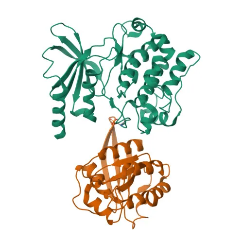
- PAK4 is a serine/threonine-protein kinase encoded by the PAK4 gene (GeneID 10298), also known as p21-activated kinase 4 (PAK-4), with a predicted molecular weight of ~64 kDa.
- Functions as a key downstream effector of the Rho family GTPases CDC42 and RAC1, regulating cytoskeletal dynamics, cell adhesion turnover, migration, growth, proliferation, and cell survival. Related references: PMID:26598620
- Upon activation, undergoes autophosphorylation and phosphorylates multiple substrates including SSH1, LIMK1, ITGB5, ARHGEF2, and BAD, thereby modulating actin filament stability, focal adhesion assembly, and apoptotic pathways. Related references: PMID:26598620
- Exerts kinase-independent effects such as preventing caspase-8 binding to death receptors and stabilizing RHOU, contributing to focal adhesion disassembly during migration. Related references: PMID:26598620
- Plays a role in cell-cycle progression by controlling CDKN1A levels and phosphorylating RAN.
- Localizes predominantly to the cytoplasm.
- Highest expression in prostate, testis, and colon.
- Belongs to the PAK family of serine/threonine-protein kinases; annotated with keywords including ATP-binding, apoptosis, cell cycle, phosphoprotein, and ubl conjugation.
Experimental Guidance and Technical Tips
- The immunogen is a synthetic peptide corresponding to the C-terminal region (aa 450-550) of human PAK4. This antibody may recognize full-length PAK4 and potentially certain alternative splicing isoforms; consider verifying isoform specificity in your system.
- For Western blot (WB), expect a band around 64 kDa in human and mouse samples. Use appropriate loading controls and validate in your specific tissue or cell lysate.
- For immunofluorescence (IF/ICC), PAK4 typically localizes to the cytoplasm; fixation and permeabilization conditions should be optimized for your cell type.
- For ELISA, pair this polyclonal antibody with the appropriate recombinant PAK4 protein or peptide antigen as a positive control.
- Cross-reactivity with human and mouse is indicated; if using other species, test for reactivity before applying to novel sample types.
CamelBio: Your One-Stop Sourcing Bridge
CamelBio provides diagnostic manufacturers, laboratories, and research institutes with a comprehensive sourcing solution for IVD raw materials, including validated antibody pairs, optimized monoclonal and polyclonal antibodies, bulk ancillary reagents, and rare target raw-material procurement. Our PAK4 antibody supports research in cell signaling, cancer biology, and cell migration, and we can assist with tailored sourcing for your specific assay development needs. Trust CamelBio as your reliable partner from concept to clinic.
REQUEST A QUOTE
Our professional team will reply to you within one business day. Please feel free to contact us!