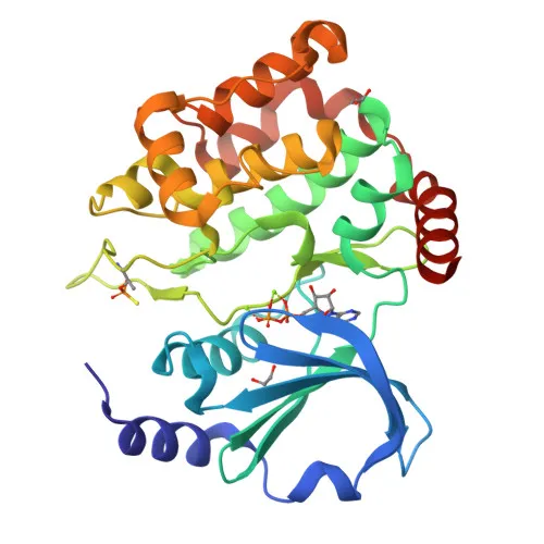
Polyclonal Antibodies
Anti-PAK3 Rabbit Polyclonal Antibody for WB, IF/ICC, ELISA - O75914
Item Number : CM0021955
Price varies based on specs and customizations
- Application
- WB, IF/ICC, ELISA
- Cross Reactivity
- Human, Mouse, Rat
- Protein Weight
- 62kDa

Shipping:
Contact us to get shipping details Enjoy On-time Dispatch Guarantee.
Why Choose Us
Easy ordering process, quality products, and dedicated support for your business success.
Core Product Specifications and Parameters
| Parameter | Value |
|---|---|
| Product Name | PAK3 Rabbit pAb |
| Remarks/Alias | ARA; bPAK; MRX30; MRX47; OPHN3; PAK-3; XLID30; PAK3beta; beta-PAK; PAK3 |
| Species | Human |
| GeneID (Human) | 5063 |
| Immunogen | Synthetic Peptide |
| Source | Rabbit |
| Category | Polyclonal Antibodies |
| Application | WB, IF/ICC, ELISA |
| Cross Reactivity | Human, Mouse, Rat |
| SWISS | O75914 |
| Protein Weight | 62kDa |
| Shipping | Ice bag |
Biological Background: PAK3 Function and Localization
- PAK3 (Serine/threonine-protein kinase PAK 3) is a member of the p21-activated kinase (PAK) family and is predominantly expressed in postmitotic neurons of the cerebral cortex and hippocampus (UniProt O75914, TISSUE SPECIFICITY).
- It functions as a downstream effector of the small GTPases CDC42 and RAC1, and plays a key role in cytoskeleton regulation, cell migration, and cell cycle regulation (FUNCTION).
- PAK3 is critical for dendrite spine morphogenesis, synapse formation, and synaptic plasticity; in hippocampal neurons, it is necessary for the formation of dendritic spines and excitatory synapses (FUNCTION, TISSUE SPECIFICITY).
- Upon activation by CDC42/RAC1, PAK3 undergoes conformational change and autophosphorylation on serine/threonine residues, then phosphorylates targets such as MAPK4, MAPK6, MAPKAPK5, and TNNI3/troponin I to regulate actin polymerization and calcium sensitivity (FUNCTION).
- Additional substrates include myosin II regulatory light chain (MLC), linking PAK3 kinase activity to actomyosin contractility in dendritic spines (FUNCTION, By similarity).
- The protein localizes to the cytoplasm (SUBCELLULAR LOCATION) and contains an SH3-binding domain and a ubiquitin-like domain (KEYWORDS: SH3-binding, Ubl conjugation).
- PAK3 is associated with X-linked intellectual disability (MRX30, MRX47, XLID30) and variant forms are linked to disease (KEYWORDS: Disease variant, Intellectual disability).
Experimental Guidance and Technical Tips
- The antibody was raised against a synthetic peptide from the N-terminal region (aa 1-100) of human PAK3 (isoform NP_001121638.1). This region may be present in multiple isoforms; specificity should be verified using isoform-specific controls.
- For Western blot, the expected molecular weight of full-length PAK3 is approximately 62 kDa; prepare lysates from relevant neuronal cell lines or brain tissues, and include appropriate positive and negative controls.
- In immunofluorescence (IF/ICC), consider testing multiple fixation/permeabilization protocols and antibody dilutions to optimize detection of endogenous PAK3, which is cytoplasmic.
- ELISA applications can be used to titer the antibody using the immunizing peptide or recombinant protein; cross-reactivity with mouse and rat proteins has been reported, but validation in the specific assay system is recommended.
- As a polyclonal antibody, lot-to-lot consistency may vary; preliminary testing with your experimental setup is advised.
CamelBio: Your One-Stop Sourcing Bridge
CamelBio offers comprehensive IVD raw material sourcing from discovery to commercialization. Our portfolio includes validated antibody pairs, monoclonal and polyclonal antibodies, bulk ancillary reagents, and rare target sourcing, serving diagnostic manufacturers and research laboratories worldwide. For neurological disease biomarkers and therapeutic targets like PAK3, we can support your assay development with customized procurement solutions.
REQUEST A QUOTE
Our professional team will reply to you within one business day. Please feel free to contact us!