Monoclonal Antibodies
Anti-PAK3 Rabbit Monoclonal Antibody for WB, IHC-P, ELISA - O75914
Item Number : CM0009610
Price varies based on specs and customizations
- Application
- WB, IHC-P, ELISA
- Cross Reactivity
- Mouse, Rat
- Protein Weight
- 62 kDa

Shipping:
Contact us to get shipping details Enjoy On-time Dispatch Guarantee.
Why Choose Us
Easy ordering process, quality products, and dedicated support for your business success.
Core Product Specifications and Parameters
| Parameter | Value |
|---|---|
| Product Name | PAK3 Rabbit mAb |
| Remarks/Alias | ARA; bPAK; MRX30; MRX47; OPHN3; PAK-3; XLID30; PAK3beta; beta-PAK; PAK3 |
| Species | Human |
| GeneID (Human) | 5063 |
| GeneID | 5063 |
| Immunogen | Synthetic peptide corresponding to a sequence within amino acids 1-100 of human PAK3 (O75914) |
| Source | Rabbit |
| Category | Monoclonal Antibodies |
| Application | WB, IHC-P, ELISA |
| Cross Reactivity | Mouse, Rat |
| SWISS | O75914 |
| Protein Weight | 62 kDa |
| Shipping | Ice bag |
Biological Background: PAK3 Function and Localization
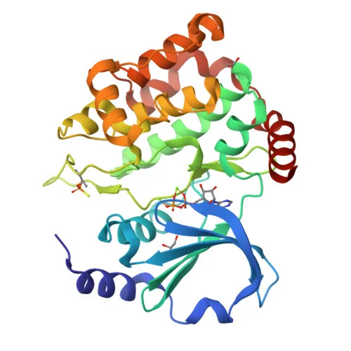
- PAK3 is a serine/threonine-protein kinase belonging to the p21-activated kinase (PAK) family.
- It acts as a downstream effector of the small GTPases CDC42 and RAC1, regulating cytoskeleton dynamics, cell migration, and cell cycle progression.
- In neurons, PAK3 plays a critical role in dendrite spine morphogenesis, synapse formation, and synaptic plasticity, partly through phosphorylation of myosin II regulatory light chain (MLC) to control actomyosin contractility.
- PAK3 is highly expressed in the nervous system, specifically in postmitotic neurons of the developing and postnatal cerebral cortex and hippocampus.
- The kinase is activated by binding to active CDC42/RAC1, leading to conformational change and autophosphorylation on multiple serine/threonine residues.
- PAK3 phosphorylates downstream targets such as MAPK4, MAPK6, and TNNI3/troponin I, linking it to MAPKAPK5-mediated F-actin polymerization and cardiac muscle contractility.
- Subcellularly, PAK3 localizes predominantly to the cytoplasm.
- Alternative splicing generates multiple isoforms; mutations in PAK3 are associated with X-linked intellectual disability.
Experimental Guidance and Technical Tips
- The immunogen is a synthetic peptide corresponding to the N-terminal region (aa 1-100) of human PAK3, suggesting the antibody likely recognizes an epitope within this domain, which may be conserved across mouse and rat.
- For Western blot (WB), the predicted band is ~62 kDa; consider using neuronal tissue or cell lysates (e.g., brain, hippocampus) as positive controls.
- For immunohistochemistry (IHC-P), paraffin-embedded brain sections are recommended; appropriate antigen retrieval methods should be optimized.
- ELISA can be performed with the immunogen peptide or recombinant PAK3 protein; cross-reactivity with related PAK family members should be assessed.
- Since PAK3 is expressed in both developing and adult nervous system, developmental stage-specific validation may be necessary.
- Always include appropriate negative controls (e.g., knockout cell lines or tissues) to confirm specificity.
CamelBio: Your One-Stop Sourcing Bridge
CamelBio empowers diagnostic manufacturers and researchers with streamlined access to high-quality IVD raw materials. For neuroscience and kinase signaling studies, we offer a curated portfolio of PAK3 antibodies, validated antibody pairs, and ancillary reagents to accelerate your assay development. Our team also specializes in sourcing rare-target raw materials, ensuring a reliable supply chain from concept to clinic.
REQUEST A QUOTE
Our professional team will reply to you within one business day. Please feel free to contact us!