Polyclonal Antibodies
Anti-PAK2 Polyclonal Antibody for WB and ELISA - Q13177
Item Number : CM0026202
Price varies based on specs and customizations
- Application
- WB, ELISA
- Cross Reactivity
- Human, Mouse, Rat
- Protein Weight
- 58kDa

Shipping:
Contact us to get shipping details Enjoy On-time Dispatch Guarantee.
Why Choose Us
Easy ordering process, quality products, and dedicated support for your business success.
Core Product Specifications and Parameters
| Parameter | Value |
|---|---|
| Product Name | PAK2 Rabbit pAb |
| Remarks/Alias | KNO2; PAK65; PAKgamma; PAK2 |
| Species | Human |
| GeneID (Human) | 5062 |
| GeneID | 5062 |
| Immunogen | Recombinant fusion protein containing a sequence corresponding to amino acids 120-240 of human PAK2 (NP_002568.2). |
| Source | Rabbit |
| Category | Polyclonal Antibodies |
| Application | WB, ELISA |
| Cross Reactivity | Human, Mouse, Rat |
| SWISS | Q13177 |
| Protein Weight | 58kDa |
| Shipping | Ice bag |
Biological Background: PAK2 Function and Localization
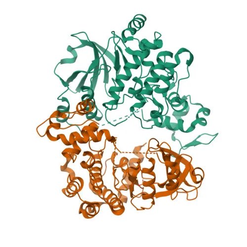
- PAK2 is a serine/threonine kinase, also known as Gamma-PAK, PAK65, and p58, encoded by the PAK2 gene.
- Acts as a downstream effector of the small GTPases CDC42 and RAC1, with activation triggering autophosphorylation on multiple serine/threonine residues.
Related references:
PMID:7744004 - Full-length PAK2 stimulates cell survival and growth, and phosphorylates MAPK4 and MAPK6 to activate MAPKAPK5, regulating F-actin polymerization and cell migration.
Related references:
PMID:7744004, PMID:21317288 - Phosphorylates diverse substrates, including histone H4 to promote nucleosome assembly, and JUN to mediate EGF-induced cell proliferation.
Related references:
PMID:21724829, PMID:21177766 - During apoptosis, caspase cleavage generates the constitutively active PAK-2p34 fragment, which translocates to the nucleus and promotes apoptosis via the JNK pathway; it also phosphorylates MKNK1 to reduce cellular translation.
Related references:
PMID:12853446, PMID:16617111, PMID:9171063, PMID:15234964 - Performs kinase-independent functions in spindle orientation during mitosis by associating with ARHGEF7 and GIT1.
Related references:
PMID:19273597, PMID:19923322 - Subcellular localization includes the cytoplasm, nucleus, perinuclear region, and membrane.
- Ubiquitously expressed with higher levels in skeletal muscle, ovary, thymus, and spleen.
- Post-translational modifications include phosphorylation, acetylation, myristoylation, and ubiquitination, which regulate its activity and localization.
Experimental Guidance and Technical Tips
- The immunogen corresponds to amino acids 120-240 within the N-terminal regulatory domain of PAK2. The resulting polyclonal antibody may detect both full-length PAK2 (~58 kDa) and the caspase-cleaved p34 fragment, depending on epitope exposure. Consider validating in both untreated and apoptotic cell lysates.
- For Western blot, expect a primary band at approximately 58 kDa; an additional ~34 kDa band may appear under apoptosis-inducing conditions.
- ELISA can be employed for quantitative detection; include recombinant PAK2 as a standard curve, and optimize coating and detection conditions.
- The antibody demonstrates cross-reactivity with mouse and rat PAK2, making it suitable for rodent model studies; however, always verify specificity in the relevant tissue or cell line.
- As a polyclonal antibody, lot-to-lot consistency may vary; include positive and negative controls in each experiment.
CamelBio: Your One-Stop Sourcing Bridge
CamelBio is a dedicated IVD raw-material partner, offering a comprehensive portfolio from monoclonal antibodies and antibody pairs to recombinant proteins and ancillary reagents. For PAK2-related kinase and apoptosis research, we supply validated products like this polyclonal antibody, alongside bulk and custom sourcing options. With technical support spanning concept to clinic, CamelBio bridges your diagnostic development needs efficiently.
REQUEST A QUOTE
Our professional team will reply to you within one business day. Please feel free to contact us!