Polyclonal Antibodies
Anti-PAK1/PAK2/PAK3 Rabbit Polyclonal Antibody for WB, ELISA - Q13153/Q13177/O75914
Item Number : CM0020046
Price varies based on specs and customizations
- Application
- WB, ELISA
- Cross Reactivity
- Human, Mouse, Rat
- Protein Weight
- 65kDa

Shipping:
Contact us to get shipping details Enjoy On-time Dispatch Guarantee.
Why Choose Us
Easy ordering process, quality products, and dedicated support for your business success.
Core Product Specifications and Parameters
| Parameter | Value |
|---|---|
| Product Name | PAK1/PAK2/PAK3 Rabbit pAb |
| Remarks/Alias | PAKalpha; PAK1/PAK2/PAK3 |
| Species | Human |
| Gene ID (Human) | 5058/5062/5063 |
| Gene ID | 5058/5062/5063 |
| Immunogen | A synthetic peptide corresponding to a sequence within amino acids 350-450 of human PAK1/PAK2/PAK3 (NP_002567.3) |
| Source | Rabbit |
| Category | Polyclonal Antibodies |
| Application | WB, ELISA |
| Cross Reactivity | Human, Mouse, Rat |
| SWISS | Q13153/Q13177/O75914 |
| Protein Weight | 65kDa |
| Shipping | Ice bag |
Biological Background: PAK1 Function and Localization
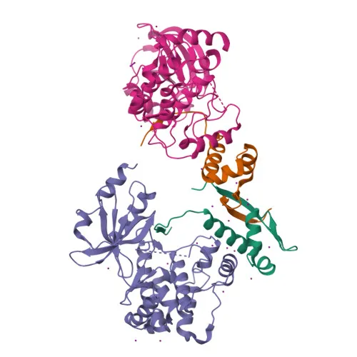
- PAK1 (p21-activated kinase 1, Alpha-PAK, p65-PAK) is a serine/threonine-protein kinase belonging to the PAK family, which also includes PAK2 (Q13177) and PAK3 (O75914). It acts as a key effector of the small GTPases CDC42 and RAC1, linking them to downstream signaling cascades. Related references: PMID:8805275 PMID:9528787
- PAK1 regulates a broad range of cellular processes, including cytoskeleton reorganization, cell adhesion, migration, proliferation, apoptosis, and mitosis. It phosphorylates numerous substrates (e.g., BAD, RAF1, MAP2K1, SNAI1, TBCB) to modulate these functions. Related references: PMID:10551809 PMID:11896197 PMID:12876277 PMID:11733498 PMID:15833848 PMID:15831477 PMID:9032240 PMID:9395435
- In the nervous system, PAK1 contributes to dendritic spine formation, synaptic plasticity, and GABA(A) receptor stability through actin filament regulation. It also phosphorylates FXR1 and MORC2, implicating it in stress granule assembly and chromatin remodeling. Related references: PMID:20417602 PMID:23260667
- Subcellularly, PAK1 localizes to the cytoplasm, focal adhesions, lamellipodia, membrane ruffles, invadopodia, nucleus, and centrosome, reflecting its dynamic roles in motility and signaling.
- PAK1 is overexpressed in gastric cancer, where it promotes cell migration and invasion, in part through RUFY3-mediated mechanisms. Related references: PMID:25766321
- Keyword annotations highlight its involvement in apoptosis, exocytosis, and disease variants (epilepsy, intellectual disability), underscoring its therapeutic relevance.
Experimental Guidance and Technical Tips
- The immunogen spans a conserved region (aa 350-450) shared across human PAK1, PAK2, and PAK3; thus, this pan‑PAK antibody likely detects all three isoforms. Consider validating specificity with isoform-specific siRNA or ectopic expression.
- For Western blot, PAK1 migrates at ~65 kDa, but PAK2 and PAK3 exhibit similar mobilities; confirm bands via parallel knockdown or phosphatase treatment where appropriate.
- ELISA protocols require empirical dilution optimization. Using recombinant PAK1/2/3 proteins or blocking peptides can help ensure signal specificity.
- Reported cross‑reactivity with mouse and rat samples should be verified in your experimental system, as post‑translational modifications or splice variants may affect detection.
CamelBio: Your One-Stop Sourcing Bridge
CamelBio is your reliable partner for IVD raw‑material sourcing, offering validated antibody pairs, optimized monoclonal and polyclonal antibodies, and bulk ancillary reagents. This anti‑PAK1/2/3 polyclonal antibody supports cytoskeleton dynamics and cell signaling research; we can also assist with rare target sourcing and custom antibody development to accelerate your diagnostic or research programs.
Product Datasheet
Anti-PAK1/PAK2/PAK3 Rabbit Polyclonal Antibody for WB, ELISA - Q13153/Q13177/O75914
REQUEST A QUOTE
Our professional team will reply to you within one business day. Please feel free to contact us!