Monoclonal Antibodies
Anti-PADI1/PAD1 Rabbit Monoclonal Antibody for WB, ELISA - Q9ULC6
Item Number : CM0007636
Price varies based on specs and customizations
- Application
- WB, ELISA
- Cross Reactivity
- Human, Mouse
- Protein Weight
- 74kDa

Shipping:
Contact us to get shipping details Enjoy On-time Dispatch Guarantee.
Why Choose Us
Easy ordering process, quality products, and dedicated support for your business success.
Core Product Specifications and Parameters
| Parameter | Value |
|---|---|
| Product Name | PADI1/PAD1 Rabbit mAb |
| Remarks/Alias | PADI1; HPAD10; PAD1; PDI; PDI1; peptidyl arginine deiminase 1; PADI1/PAD1 |
| Species | Human |
| Gene ID (Human) | 29943 |
| Gene ID | 29943 |
| Immunogen | Recombinant fusion protein containing a sequence corresponding to amino acids 1-277 of human PADI1/PAD1 (NP_037490.2) |
| Source | Rabbit |
| Category | Monoclonal Antibodies |
| Application | WB, ELISA |
| Cross Reactivity | Human, Mouse |
| SWISS | Q9ULC6 |
| Protein Weight | 74kDa |
| Shipping | Ice bag |
Biological Background: PADI1/PAD1 Function and Localization
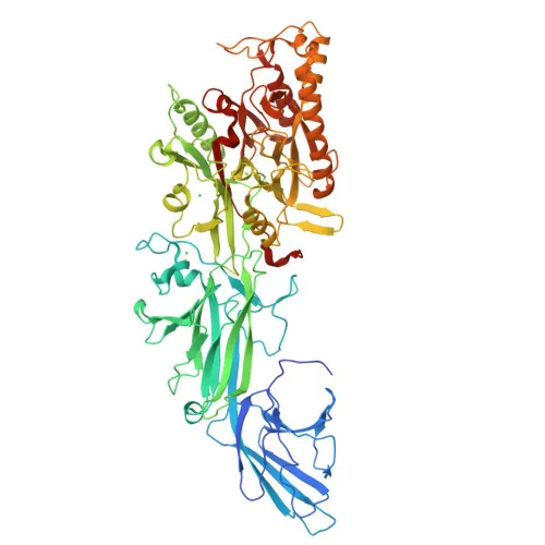
- Protein-arginine deiminase type-1 (PADI1), also known as peptidylarginine deiminase I, is encoded by the PADI1 gene (alternative symbols: PAD1, PDI1).
- Catalyzes the deimination (citrullination) of arginine residues in target proteins, converting positively charged arginine to neutral citrulline.
- Localizes to the cytoplasm, as annotated in UniProt, where it is expected to interact with its substrate proteins.
- Expressed in a tissue-specific manner: highly detected in epidermal keratinocytes and also found in prostate, testis, placenta, spleen, and thymus, suggesting roles in skin biology and other tissues.
- Classified as a hydrolase and a metal-binding protein requiring calcium for activity; its 3D structure has been determined.
- Involved in post-translational modification pathways that can alter protein function and antigenicity.
Experimental Guidance and Technical Tips
- The immunogen comprises the N-terminal 277 amino acids of human PADI1, which may allow detection of both full-length and N-terminal fragments; consider using positive controls expressing full-length protein.
- For Western blot (WB), validate the antibody with a dilution series under reducing conditions and confirm band specificity using lysates from known PADI1-expressing cells or tissues (e.g., keratinocytes).
- In ELISA applications, coat recombinant PADI1 protein or cell lysates and titrate the antibody to determine optimal coating and detection concentrations.
- Given reported cross-reactivity with mouse, this antibody is suitable for translational studies in mouse models; always confirm cross-reactivity experimentally in your system.
CamelBio: Your One-Stop Sourcing Bridge
CamelBio is your global one-stop partner for IVD raw materials, offering comprehensive access to validated monoclonal antibodies like this PADI1/PAD1 clone. Whether you need bulk quantities, ancillary reagents, or assistance in sourcing rare targets, our platform streamlines procurement from concept to clinic. Let us support your next project in protein citrullination research with reliable, high-quality raw materials.
REQUEST A QUOTE
Our professional team will reply to you within one business day. Please feel free to contact us!