Polyclonal Antibodies
Anti-PACSIN2 Rabbit Polyclonal Antibody for WB, ELISA - Q9UNF0
Item Number : CM0022742
Price varies based on specs and customizations
- Application
- WB, ELISA
- Cross Reactivity
- Human, Mouse
- Protein Weight
- 56kDa

Shipping:
Contact us to get shipping details Enjoy On-time Dispatch Guarantee.
Why Choose Us
Easy ordering process, quality products, and dedicated support for your business success.
Core Product Specifications and Parameters
| Parameter | Value |
|---|---|
| Product Name | PACSIN2 Rabbit pAb |
| Remarks/Alias | SDPII; PACSIN2 |
| Species | Human |
| GeneID (Human) | 11252 |
| GeneID | 11252 |
| Immunogen | Recombinant fusion protein containing a sequence corresponding to amino acids 240-410 of human PACSIN2 (NP_009160.2) |
| Source | Rabbit |
| Category | Polyclonal Antibodies |
| Application | WB, ELISA |
| Cross Reactivity | Human, Mouse |
| SWISS | Q9UNF0 |
| Protein Weight | 56kDa |
| Shipping | Ice bag |
Biological Background: PACSIN2 Function and Localization
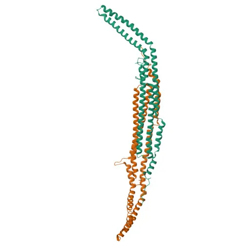
- PACSIN2 (Syndapin-2) is a lipid-binding protein that promotes tubulation of phosphatidic acid-containing membranes and plays a key role in intracellular vesicle-mediated transport, including caveolae morphogenesis and endocytosis (By similarity).
- It is involved in the endocytosis of cell-surface receptors such as the EGF receptor, contributing to its internalization even in the absence of EGF stimulus. Related references: PMID:21693584, PMID:23129763, PMID:23236520
- In microbial infection contexts, PACSIN2 enhances HIV-1 cell-to-cell spread and promotes protrusion engulfment during bacterial spread (e.g., Listeria monocytogenes), and is involved in lipid droplet formation important for HCV virion assembly. Related references: PMID:29891700, PMID:31242077, PMID:31801866
- Subcellularly, PACSIN2 localizes to the cytoplasm, cytoskeleton, cytoplasmic vesicle membranes, cell projections (ruffles and caveolae), early and recycling endosomes, cell membrane, and adherens junctions.
- It is essential for endothelial organization in sprouting angiogenesis by modulating CDH5-based junctions and facilitating front-rear polarity through recruitment of EHD4 and MICALL1 (By similarity).
- The protein contains a coiled-coil region and an SH3 domain, and is subject to acetylation and phosphorylation, as indicated by proteomic identification data.
- PACSIN2 is widely expressed in tissues, with no reported tissue-specific limitations.
Experimental Guidance and Technical Tips
- The immunogen corresponds to amino acids 240-410 of human PACSIN2 (NP_009160.2), a region likely to recognize both human and mouse PACSIN2 given the cross-reactivity profile.
- For Western blot (WB), a band around 56 kDa is expected; consider using appropriate positive controls such as human or mouse cell lysates expressing PACSIN2.
- In ELISA applications, the antibody may be used for detection of recombinant or endogenous PACSIN2; validate under the desired experimental conditions.
- Since PACSIN2 is widely expressed and localizes to multiple subcellular compartments, consider fractionation or immunostaining protocols to study its specific localization.
CamelBio: Your One-Stop Sourcing Bridge
CamelBio provides a reliable one-stop supply chain for IVD raw materials, including validated antibody pairs, optimized polyclonal antibodies, and bulk ancillary reagents. For targets like PACSIN2, which plays critical roles in cell trafficking and infectious disease pathways, CamelBio can source rare raw materials and assist in assay development. Count on CamelBio to streamline your procurement from concept to clinic.
REQUEST A QUOTE
Our professional team will reply to you within one business day. Please feel free to contact us!