Monoclonal Antibodies
Anti-p73 Rabbit Monoclonal Antibody for WB, IHC-P, ELISA - O15350
Item Number : CM0006984
Price varies based on specs and customizations
- Application
- WB, IHC-P, ELISA
- Cross Reactivity
- Human, Mouse, Rat
- Protein Weight
- 70kDa

Shipping:
Contact us to get shipping details Enjoy On-time Dispatch Guarantee.
Why Choose Us
Easy ordering process, quality products, and dedicated support for your business success.
Core Product Specifications and Parameters
| Parameter | Value |
|---|---|
| Product Name | p73 Rabbit mAb |
| Remarks/Alias | P73; CILD47; p73 |
| Species | Human |
| Gene ID | 7161 |
| Immunogen | A synthetic peptide corresponding to a sequence within amino acids 1-100 of human p73 (O15350). |
| Source | Rabbit |
| Category | Monoclonal Antibodies |
| Application | WB, IHC-P, ELISA |
| Cross Reactivity | Human, Mouse, Rat |
| SWISS | O15350 |
| Protein Weight | 70kDa |
| Shipping | Ice bag |
Biological Background: p73 Function and Localization
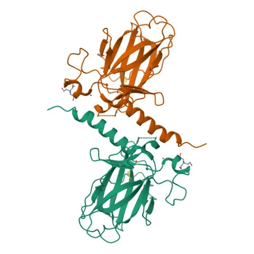
- Protein Identity and Isoforms: Tumor protein p73, also known as p53-like transcription factor and p53-related protein, belongs to the p53 family. Multiple isoforms exist, differing in the presence of the N-terminal transactivation domain.
- Apoptotic Response and Tumor Suppression: p73 participates in the apoptotic response to DNA damage. Isoforms containing the transactivation domain are pro-apoptotic, while isoforms lacking this domain are anti-apoptotic and can inhibit p53 and transactivating p73 isoforms. p73 may function as a tumor suppressor.
- Ciliated Cell Differentiation: p73 is an essential factor for the positive regulation of lung ciliated cell differentiation, acting as an activator of FOXJ1 expression. Related references: PMID:34077761
- Subcellular Localization: p73 localizes to both the nucleus and the cytoplasm.
- Tissue Expression: p73 is expressed in a wide range of tissues including brain, kidney, placenta, colon, heart, liver, spleen, skeletal muscle, prostate, thymus, and pancreas. It is highly expressed in fetal tissue and is found in the respiratory epithelium. At the protein level, it is also detected in striatal neurons of patients with Huntington disease.
- Keywords and Functional Features: p73 is a DNA-binding transcription factor involved in apoptosis, cell cycle regulation, and transcription. It is a phosphoprotein, metal-binding, and subject to ubiquitin-like conjugation. Mutations are associated with ciliopathy and lissencephaly.
Experimental Guidance and Technical Tips
- The antibody was generated against an N-terminal synthetic peptide (aa 1–100) of human p73. For cross-reactivity with mouse and rat, verify sequence homology in this region.
- In Western blotting (WB), a band at approximately 70 kDa is expected. Use appropriate molecular weight markers and validate with positive and negative controls.
- For immunohistochemistry on paraffin-embedded sections (IHC-P), optimizing antigen retrieval and antibody dilution is recommended. Include tissue controls known to express p73.
- The antibody is suitable for ELISA applications; use purified recombinant protein or peptide for standard curve generation.
CamelBio: Your One-Stop Sourcing Bridge
CamelBio provides diagnostic manufacturers, labs, and research institutes with one-stop access to IVD raw materials, technical services, and consulting—covering every stage from concept to clinic. This Anti-p73 Rabbit Monoclonal Antibody supports apoptosis and cancer research applications. As your sourcing partner, CamelBio can supply validated antibody pairs, optimized monoclonal antibodies, bulk ancillary reagents, and assist with rare target raw-material procurement to accelerate your diagnostic development.
REQUEST A QUOTE
Our professional team will reply to you within one business day. Please feel free to contact us!