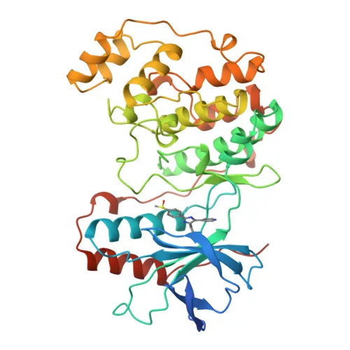
Monoclonal Antibodies
Anti-p38 MAPK Rabbit Monoclonal Antibody for WB, IF/ICC, ELISA - Q16539
Item Number : CM0005186
Price varies based on specs and customizations
- Application
- WB, IF/ICC, ELISA
- Cross Reactivity
- Human, Mouse, Rat
- Protein Weight
- 41kDa

Shipping:
Contact us to get shipping details Enjoy On-time Dispatch Guarantee.
Why Choose Us
Easy ordering process, quality products, and dedicated support for your business success.
Core Product Specifications and Parameters
| Parameter | Value |
|---|---|
| Product Name | p38 MAPK Rabbit mAb |
| Remarks/Alias | RK; p38; CSBP; EXIP; Mxi2; CSBP1; CSBP2; CSPB1; PRKM14; PRKM15; SAPK2A; p38ALPHA; p38 MAPK |
| Species | Human |
| Gene ID | 1432 |
| Immunogen | Synthetic Peptide; A synthetic peptide corresponding to a sequence within amino acids 100-200 of human p38 MAPK (Q16539) |
| Source | Rabbit |
| Category | Monoclonal Antibodies |
| Application | WB, IF/ICC, ELISA |
| Cross Reactivity | Human, Mouse, Rat |
| SWISS | Q16539 |
| Protein Weight | 41kDa |
| Shipping | Ice bag |
Biological Background: p38 MAPK (MAPK14) Function and Localization
- MAPK14 is a serine/threonine kinase that serves as a core component of the p38 MAP kinase pathway. Activated by pro-inflammatory cytokines and various stressors, it regulates an extensive network of downstream substrates (estimated 200–300 targets) to control diverse cellular processes including gene expression, mRNA stability, translation, and protein turnover.
- Through phosphorylation of downstream kinases MSK1/2 and MAPKAPK2/3/MNK1/2, MAPK14 orchestrates transcriptional and post‑transcriptional regulation. MSK1/2 activate transcription factors (CREB1, ATF1, NF‑κB, STAT1/3) and directly phosphorylate histone H3 and HMGN1 to promote chromatin remodeling and immediate‑early gene induction. MAPKAPK2/3 control mRNA stability via ZFP36 and ELAVL1, while MNK1/2 modulate translation through eIF4E2 phosphorylation. Related references: PMID:9687510, PMID:9792677, PMID:11154262
- MAPK14 directly phosphorylates a range of transcription factors, including TP53/p53 (in cooperation with casein kinase II), ATF1, ATF2, ELK1, and MEF2C/MEF2A, linking stress signals to transcriptional reprogramming. Related references: PMID:10747897, PMID:10330143, PMID:9430721, PMID:9858528
- The kinase regulates protein degradation and membrane receptor dynamics. It phosphorylates CFLAR to promote proteasomal degradation, controls SIAH2 ubiquitin ligase activity, and interferes with ATG9 trafficking to modulate autophagy. Moreover, MAPK14 phosphorylates EGFR and RAB5A effectors to regulate clathrin‑mediated endocytosis, and activates ADAM17‑driven ectodomain shedding of EGFR ligands. Related references: PMID:17003045, PMID:19893488, PMID:16932740, PMID:20188673
- MAPK14 participates in cell cycle checkpoints and DNA damage responses. It phosphorylates CDC25B and CDC25C, enabling 14‑3‑3 binding and initiating a G2 delay after UV radiation. Upon DNA damage, it phosphorylates TIAR, which releases TIAR from GADD45A mRNA and prevents transcript degradation. Related references: PMID:11333986, PMID:20932473
- Essential for placental labyrinth blood vessel growth and stress‑induced erythropoiesis through regulation of EPO gene expression. MAPK14 also triggers NLRP1 inflammasome activation downstream of ribosome collisions, leading to pyroptosis. Additionally, during M. tuberculosis infection, bacterial EsxA phosphorylates MAPK14 to suppress IFN‑γ production in T cells. Related references: PMID:10943842, PMID:35857590, PMID:21586573
- MAPK14 is expressed in a wide range of tissues, with highest levels in brain, heart, placenta, pancreas, and skeletal muscle; lower expression is observed in lung, liver, and kidney. It localizes to both cytoplasm and nucleus. Alternative splicing produces isoforms such as MXI2 (reduced kinase activity) and EXIP (pro‑apoptotic). The protein undergoes multiple post‑translational modifications including phosphorylation, acetylation, and ubiquitin‑like conjugation.
Experimental Guidance and Technical Tips
- The immunogen (synthetic peptide mapping to amino acids 100‑200) lies within a conserved region; thus cross‑reactivity with mouse and rat MAPK14 is expected. Validate reactivity in relevant species‑specific cell or tissue lysates before routine use.
- For western blot, consider using whole‑cell extracts from stressed vs. resting cells to observe phospho‑specific band shifts. Always include appropriate controls (e.g., phospho‑p38 antibodies, kinase inhibitors) to ensure signal specificity.
- In immunofluorescence/ICC, optimize fixation/permeabilization conditions and confirm staining patterns consistent with cytoplasmic and nuclear distribution. Co‑staining with compartment markers is recommended.
- For ELISA, use recombinant human MAPK14 protein as a standard and validate linearity of detection. Pair this antibody with a suitable detection antibody to develop a sensitive sandwich ELISA.
- Since MAPK14 exhibits kinase‑independent functions and multiple isoforms, verify that your experimental readout reflects the specific role you intend to study.
CamelBio: Your One-Stop Sourcing Bridge
CamelBio offers diagnostic developers, research institutes, and laboratories a streamlined sourcing solution for IVD raw materials, from monoclonal and polyclonal antibodies to bulk reagents and technical consulting. For projects focused on p38 MAPK signaling, inflammation, or stress‑response pathways, we provide consistent access to validated antibodies like this anti‑p38 MAPK rabbit monoclonal antibody, along with the capability to source rare‑target materials, enabling you to advance from early‑stage research to clinical application efficiently.
Product Datasheet
Anti-p38 MAPK Rabbit Monoclonal Antibody for WB, IF/ICC, ELISA - Q16539
REQUEST A QUOTE
Our professional team will reply to you within one business day. Please feel free to contact us!