Polyclonal Antibodies
Anti-OPA1 Rabbit Polyclonal Antibody for WB, IF/ICC, IHC-P, ELISA - O60313
Item Number : CM0025167
Price varies based on specs and customizations
- Application
- WB, IF/ICC, IHC-P, ELISA
- Cross Reactivity
- Human, Mouse, Rat
- Protein Weight
- 112kDa

Shipping:
Contact us to get shipping details Enjoy On-time Dispatch Guarantee.
Why Choose Us
Easy ordering process, quality products, and dedicated support for your business success.
Core Product Specifications and Parameters
| Parameter | Value |
|---|---|
| Product Name | OPA1 Rabbit pAb |
| Remarks/Alias | NPG; NTG; MGM1; BERHS; largeG; MTDPS14; OPA1 |
| Species | Human |
| GeneID (human) | 4976 |
| GeneID | 4976 |
| Immunogen | Recombinant fusion protein containing a sequence corresponding to amino acids 200-600 of human OPA1 (NP_056375.2) |
| Source | Rabbit |
| Category | Polyclonal Antibodies |
| Application | WB, IF/ICC, IHC-P, ELISA |
| Cross Reactivity | Human, Mouse, Rat |
| SWISS | O60313 |
| Protein Weight | 112kDa |
| Shipping | Ice bag |
Biological Background: OPA1 Function and Localization
- OPA1 (Optic atrophy protein 1) is a dynamin-like GTPase that exists as two main forms: a long transmembrane form (L-OPA1) tethered to the inner mitochondrial membrane, and a short soluble form (S-OPA1) in the intermembrane space, generated by OMA1 cleavage (PubMed:31922487, PubMed:32228866, PubMed:33237841). Related references: PMID:31922487, PMID:32228866, PMID:33237841
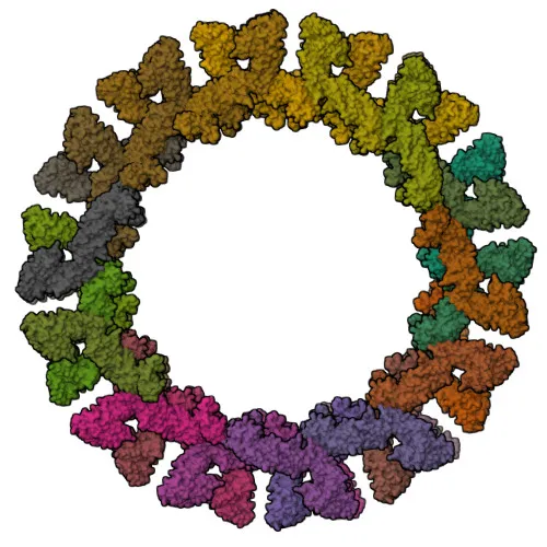
- OPA1 is a key mediator of mitochondrial inner membrane fusion and cristae morphology. L-OPA1 and S-OPA1 cooperate to form helical assemblies that drive membrane curvature and fusion via GTP hydrolysis (PubMed:28628083, PubMed:37612504, PubMed:37612506). Related references: PMID:28628083, PMID:37612504, PMID:37612506
- Belongs to the dynamin superfamily of large GTPases, characterized by conserved domains involved in self-assembly and membrane remodeling.
- Subcellular location: Mitochondrion inner membrane and intermembrane space.
- Essential for mitochondrial genome maintenance (PubMed:18158317, PubMed:20974897) and regulation of apoptosis through cristae remodeling and cytochrome c release. Related references: PMID:18158317, PMID:20974897
- Highly expressed in retina, brain, testis, heart, and skeletal muscle (PubMed:11017079, PubMed:11017080, PubMed:11810270). Related references: PMID:11017079, PMID:11017080, PMID:11810270
- Post-translational modifications: Proteolytic processing by PARL and OMA1 generates S-OPA1, influencing mitochondrial dynamics. Acetylation and disulfide bond formation are also noted.
- Disease relevance: Mutations in OPA1 cause autosomal dominant optic atrophy, and are associated with mitochondrial syndromes, deafness, and cardiomyopathy.
Experimental Guidance and Technical Tips
- For Western blot (WB), the expected molecular weight of OPA1 is approximately 112 kDa; multiple isoforms may be detected due to alternative splicing and proteolytic processing. Consider loading sufficient protein and using gradient gels for resolution.
- For immunofluorescence (IF/ICC) and immunohistochemistry (IHC-P), OPA1 localizes to mitochondria, showing a characteristic punctate pattern. Mitochondrial co-staining is recommended for verification.
- The immunogen corresponds to amino acids 200-600 of human OPA1, an internal region conserved across isoforms; thus, the antibody may recognize multiple OPA1 variants. Validate in the relevant species and sample system.
- As a polyclonal antibody, lot-to-lot variability may occur; include appropriate positive and negative controls in each experiment.
CamelBio: Your One-Stop Sourcing Bridge
CamelBio is a one-stop IVD raw-material sourcing partner for diagnostic manufacturers, labs, and research institutes. This anti-OPA1 polyclonal antibody supports research into mitochondrial dynamics, apoptosis, and optic atrophy. Beyond single antibodies, CamelBio offers validated antibody pairs, optimized monoclonal/polyclonal antibodies, bulk ancillary reagents, and custom sourcing for rare targets, helping to streamline your assay development from concept to clinic.
Product Datasheet
Anti-OPA1 Rabbit Polyclonal Antibody for WB, IF/ICC, IHC-P, ELISA - O60313
REQUEST A QUOTE
Our professional team will reply to you within one business day. Please feel free to contact us!