Polyclonal Antibodies
Anti-OLA1 Polyclonal Antibody for WB, IHC-P, IF/ICC, ELISA - Q9NTK5
Item Number : CM0016822
Price varies based on specs and customizations
- Application
- WB, IHC-P, IF/ICC, ELISA
- Cross Reactivity
- Human, Mouse, Rat
- Protein Weight
- 45kDa

Shipping:
Contact us to get shipping details Enjoy On-time Dispatch Guarantee.
Why Choose Us
Easy ordering process, quality products, and dedicated support for your business success.
Core Product Specifications and Parameters
| Parameter | Value |
|---|---|
| Product Name | OLA1 Rabbit pAb |
| Remarks/Alias | DOC45, GBP45, GTBP9, GTPBP9, PTD004, OLA1 |
| Species | Human |
| GeneID | 29789 |
| Immunogen | Recombinant fusion protein containing a sequence corresponding to amino acids 130-310 of human OLA1 (NP_037473.3). |
| Source | Rabbit |
| Category | Polyclonal Antibodies |
| Application | WB, IHC-P, IF/ICC, ELISA |
| Cross Reactivity | Human, Mouse, Rat |
| SWISS | Q9NTK5 |
| Protein Weight | 45kDa |
| Shipping | Ice bag |
Biological Background: OLA1 Function and Localization
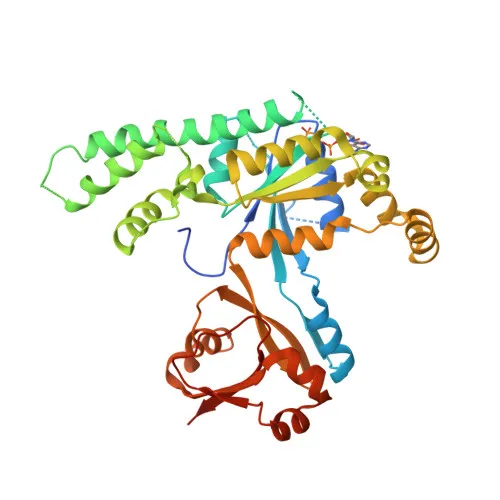
- OLA1 (Obg-like ATPase 1), also known as DOC45 or GTPBP9, is a member of the Obg family of GTPases, characterized by a conserved GTP-binding domain.
- It primarily functions as an ATPase, hydrolyzing ATP with higher efficiency than GTP, and plays a role in cellular stress responses and DNA damage repair pathways.
- The protein is expressed in various tissues, with higher abundance in testis, liver, lung, and brain, and is overexpressed in several malignancies including colon, rectal, ovarian, lung, stomach, and uterine cancers.
- Subcellularly, OLA1 localizes to the cytoplasm and nucleus, including the nucleolus, indicating involvement in both cytosolic and nuclear processes.
- Post-translational modifications include acetylation, and it binds magnesium as a cofactor for its hydrolase activity.
- Alternative splicing generates multiple transcript variants, contributing to functional diversity.
Experimental Guidance and Technical Tips
- The immunogen used is a recombinant fragment spanning amino acids 130-310, which lies within the central region of OLA1. This design may provide better accessibility to epitopes in both native and denatured states, supporting applications such as Western blotting and immunohistochemistry.
- For Western blotting, expect a band around 45 kDa. Consider using appropriate loading controls and validate the antibody in the relevant cell or tissue lysates.
- In immunohistochemistry (IHC-P), antigen retrieval conditions may need optimization due to the nuclear and cytoplasmic localization of OLA1. Perform pilot studies to determine optimal staining parameters.
- For immunofluorescence (IF/ICC), fixation and permeabilization should preserve both cytoplasmic and nuclear structures; including a nuclear counterstain can help confirm localization.
- This antibody also works in ELISA, making it suitable for screening and quantification assays; validate in the specific sample matrix of interest.
CamelBio: Your One-Stop Sourcing Bridge
CamelBio is a dedicated IVD raw-material supplier providing a comprehensive range of validated antibodies, including polyclonal and monoclonal antibodies, for diagnostic and research applications. Our portfolio supports cancer biology and cellular stress research, with OLA1 being a key target in oncological studies. From bulk ancillary reagents to specialty antibodies, we help streamline your procurement process, offering reliable sourcing from concept to clinic.
REQUEST A QUOTE
Our professional team will reply to you within one business day. Please feel free to contact us!